Conjunctiva
(Redirected from Conjunctival fornix)
Conjunctiva[edit | edit source]
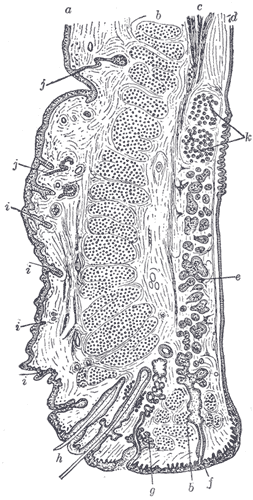
The conjunctiva is a thin, transparent mucous membrane that covers the sclera (the white part of the eye) and lines the inside of the eyelids. It plays a crucial role in maintaining the health of the eye by providing a protective barrier against environmental irritants and pathogens.
Anatomy[edit | edit source]
The conjunctiva is divided into three parts:
- Palpebral conjunctiva: This part lines the inside of the eyelids. It is highly vascularized and adheres tightly to the tarsal plates of the eyelids.
- Bulbar conjunctiva: This part covers the anterior surface of the sclera, up to the cornea. It is loosely attached to the underlying tissue, allowing for free movement of the eyeball.
- Fornix conjunctiva: This is the loose, flexible fold that connects the palpebral and bulbar conjunctiva, allowing for the movement of the eye and eyelids.
Function[edit | edit source]
The primary functions of the conjunctiva include:
- Protection: It acts as a barrier to dust, debris, and microorganisms, preventing them from entering the eye.
- Lubrication: The conjunctiva produces mucus and tears, which help to keep the eye moist and facilitate smooth movement of the eyelids over the eyeball.
- Immune defense: It contains immune cells that help to detect and respond to pathogens.
Clinical Significance[edit | edit source]
The conjunctiva can be affected by various conditions, including:
- Conjunctivitis: Also known as "pink eye," this is an inflammation of the conjunctiva, often caused by infections, allergies, or irritants.
- Pterygium: A benign growth of the conjunctiva that can extend onto the cornea, potentially affecting vision.
- Pinguecula: A yellowish, benign growth on the conjunctiva, usually on the side closest to the nose.
Histology[edit | edit source]
The conjunctiva is composed of a non-keratinized stratified columnar epithelium with goblet cells interspersed throughout. These goblet cells are responsible for secreting mucus, which contributes to the tear film and helps maintain ocular surface health.
Related Pages[edit | edit source]
See Also[edit | edit source]
Search WikiMD
Ad.Tired of being Overweight? Try W8MD's physician weight loss program.
Semaglutide (Ozempic / Wegovy and Tirzepatide (Mounjaro / Zepbound) available.
Advertise on WikiMD
|
WikiMD's Wellness Encyclopedia |
| Let Food Be Thy Medicine Medicine Thy Food - Hippocrates |
Translate this page: - East Asian
中文,
日本,
한국어,
South Asian
हिन्दी,
தமிழ்,
తెలుగు,
Urdu,
ಕನ್ನಡ,
Southeast Asian
Indonesian,
Vietnamese,
Thai,
မြန်မာဘာသာ,
বাংলা
European
español,
Deutsch,
français,
Greek,
português do Brasil,
polski,
română,
русский,
Nederlands,
norsk,
svenska,
suomi,
Italian
Middle Eastern & African
عربى,
Turkish,
Persian,
Hebrew,
Afrikaans,
isiZulu,
Kiswahili,
Other
Bulgarian,
Hungarian,
Czech,
Swedish,
മലയാളം,
मराठी,
ਪੰਜਾਬੀ,
ગુજરાતી,
Portuguese,
Ukrainian
Medical Disclaimer: WikiMD is not a substitute for professional medical advice. The information on WikiMD is provided as an information resource only, may be incorrect, outdated or misleading, and is not to be used or relied on for any diagnostic or treatment purposes. Please consult your health care provider before making any healthcare decisions or for guidance about a specific medical condition. WikiMD expressly disclaims responsibility, and shall have no liability, for any damages, loss, injury, or liability whatsoever suffered as a result of your reliance on the information contained in this site. By visiting this site you agree to the foregoing terms and conditions, which may from time to time be changed or supplemented by WikiMD. If you do not agree to the foregoing terms and conditions, you should not enter or use this site. See full disclaimer.
Credits:Most images are courtesy of Wikimedia commons, and templates, categories Wikipedia, licensed under CC BY SA or similar.
Contributors: Prab R. Tumpati, MD


