Trabecular meshwork
A part of the eye involved in draining aqueous humor
Trabecular meshwork[edit]
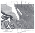
The trabecular meshwork is a crucial structure in the eye that plays a significant role in the drainage of aqueous humor, the clear fluid filling the space in the front of the eyeball between the lens and the cornea. It is located in the anterior chamber angle, where the cornea meets the iris.

Anatomy[edit]
The trabecular meshwork is a spongy tissue that forms a mesh-like structure. It is situated circumferentially around the base of the cornea, near the scleral spur. The meshwork is divided into three regions: the uveal meshwork, the corneoscleral meshwork, and the juxtacanalicular tissue. Each of these regions has distinct structural and functional properties that contribute to the regulation of aqueous humor outflow.

Function[edit]
The primary function of the trabecular meshwork is to facilitate the drainage of aqueous humor from the eye into the Schlemm's canal, and subsequently into the bloodstream. This process is essential for maintaining intraocular pressure (IOP) within a normal range. Dysfunctions in the trabecular meshwork can lead to increased IOP, which is a major risk factor for glaucoma, a group of eye conditions that can cause blindness.
Clinical significance[edit]
Abnormalities in the trabecular meshwork can lead to various eye conditions, most notably glaucoma. In open-angle glaucoma, the trabecular meshwork becomes less efficient at draining aqueous humor, leading to increased IOP. Treatments for glaucoma often aim to improve the outflow of aqueous humor through the trabecular meshwork, either through medication or surgical procedures.

Research and developments[edit]
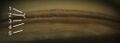
Recent advances in imaging technologies, such as optical coherence tomography (OCT), have allowed for more detailed visualization of the trabecular meshwork. These technologies aid in the diagnosis and management of glaucoma by providing high-resolution images of the anterior chamber angle and the trabecular meshwork.
.png)
Related pages[edit]
References[edit]
- Quigley, H. A. (2011). "Glaucoma." The Lancet, 377(9774), 1367-1377.
- Johnson, M. (2006). "What controls aqueous humour outflow?" Experimental Eye Research, 82(4), 545-557.
Trabecular_meshwork[edit]
-
 Trabecular meshwork illustration
 -
 Kammerwinkel
 -
 Anterior chamber angle - 3D motion parallax
 -
 SD OCT - Anterior Chamber Angle Cross-Section

Medical Disclaimer: WikiMD is for informational purposes only and is not a substitute for professional medical advice. Content may be inaccurate or outdated and should not be used for diagnosis or treatment. Always consult your healthcare provider for medical decisions. Verify information with trusted sources such as CDC.gov and NIH.gov. By using this site, you agree that WikiMD is not liable for any outcomes related to its content. See full disclaimer.
Credits:Most images are courtesy of Wikimedia commons, and templates, categories Wikipedia, licensed under CC BY SA or similar.
Translate page: - East Asian
中文,
日本,
한국어,
South Asian
हिन्दी,
தமிழ்,
తెలుగు,
Urdu,
ಕನ್ನಡ,
Southeast Asian
Indonesian,
Vietnamese,
Thai,
မြန်မာဘာသာ,
বাংলা
European
español,
Deutsch,
français,
Greek,
português do Brasil,
polski,
română,
русский,
Nederlands,
norsk,
svenska,
suomi,
Italian
Middle Eastern & African
عربى,
Turkish,
Persian,
Hebrew,
Afrikaans,
isiZulu,
Kiswahili,
Other
Bulgarian,
Hungarian,
Czech,
Swedish,
മലയാളം,
मराठी,
ਪੰਜਾਬੀ,
ગુજરાતી,
Portuguese,
Ukrainian
