Internal globus pallidus
(Redirected from Globus pallidus medialis)
Internal globus pallidus
The internal globus pallidus (GPi) is a subcomponent of the globus pallidus, which is part of the basal ganglia in the brain. The basal ganglia are a group of nuclei in the brain associated with a variety of functions including the regulation of voluntary motor movements, procedural learning, routine behaviors or "habits", eye movements, cognition, and emotion.
Anatomy[edit | edit source]
The internal globus pallidus is located within the telencephalon and is one of the two parts of the globus pallidus, the other being the external globus pallidus (GPe). The GPi is situated medial to the GPe and lateral to the thalamus. It is separated from the thalamus by the internal capsule, a white matter structure that contains many of the brain's ascending and descending axons.
Function[edit | edit source]
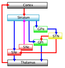
The GPi plays a crucial role in the regulation of voluntary movement. It is part of the indirect pathway and the direct pathway of the basal ganglia circuitry. In the direct pathway, the GPi receives inhibitory input from the striatum and sends inhibitory output to the thalamus, which in turn projects to the motor cortex. This pathway facilitates the initiation and execution of voluntary movements. In the indirect pathway, the GPi receives excitatory input from the subthalamic nucleus and sends inhibitory output to the thalamus, which helps to suppress unwanted movements.
Clinical Significance[edit | edit source]
Dysfunction of the GPi is associated with several movement disorders. For example, in Parkinson's disease, there is excessive inhibitory output from the GPi to the thalamus, leading to the characteristic motor symptoms of bradykinesia, rigidity, and tremor. Deep brain stimulation (DBS) of the GPi is a therapeutic option for patients with Parkinson's disease and other movement disorders such as dystonia.
Connections[edit | edit source]
The GPi has extensive connections with other parts of the basal ganglia, including the putamen, caudate nucleus, and the substantia nigra. It also has connections with the cerebral cortex and the brainstem, which are important for its role in motor control.
See Also[edit | edit source]
- Basal ganglia
- External globus pallidus
- Subthalamic nucleus
- Parkinson's disease
- Deep brain stimulation
References[edit | edit source]
This article is a neuroanatomy stub. You can help WikiMD by expanding it!
Search WikiMD
Ad.Tired of being Overweight? Try W8MD's physician weight loss program.
Semaglutide (Ozempic / Wegovy and Tirzepatide (Mounjaro / Zepbound) available.
Advertise on WikiMD
|
WikiMD's Wellness Encyclopedia |
| Let Food Be Thy Medicine Medicine Thy Food - Hippocrates |
Translate this page: - East Asian
中文,
日本,
한국어,
South Asian
हिन्दी,
தமிழ்,
తెలుగు,
Urdu,
ಕನ್ನಡ,
Southeast Asian
Indonesian,
Vietnamese,
Thai,
မြန်မာဘာသာ,
বাংলা
European
español,
Deutsch,
français,
Greek,
português do Brasil,
polski,
română,
русский,
Nederlands,
norsk,
svenska,
suomi,
Italian
Middle Eastern & African
عربى,
Turkish,
Persian,
Hebrew,
Afrikaans,
isiZulu,
Kiswahili,
Other
Bulgarian,
Hungarian,
Czech,
Swedish,
മലയാളം,
मराठी,
ਪੰਜਾਬੀ,
ગુજરાતી,
Portuguese,
Ukrainian
Medical Disclaimer: WikiMD is not a substitute for professional medical advice. The information on WikiMD is provided as an information resource only, may be incorrect, outdated or misleading, and is not to be used or relied on for any diagnostic or treatment purposes. Please consult your health care provider before making any healthcare decisions or for guidance about a specific medical condition. WikiMD expressly disclaims responsibility, and shall have no liability, for any damages, loss, injury, or liability whatsoever suffered as a result of your reliance on the information contained in this site. By visiting this site you agree to the foregoing terms and conditions, which may from time to time be changed or supplemented by WikiMD. If you do not agree to the foregoing terms and conditions, you should not enter or use this site. See full disclaimer.
Credits:Most images are courtesy of Wikimedia commons, and templates, categories Wikipedia, licensed under CC BY SA or similar.
Contributors: Prab R. Tumpati, MD


