Inferior extensor retinaculum of the foot
A band of connective tissue in the foot
The inferior extensor retinaculum of the foot is a crucial anatomical structure that plays a significant role in the stability and function of the foot. It is a thickened band of connective tissue located on the anterior aspect of the ankle and foot, serving to hold the tendons of the extensor muscles in place as they pass over the ankle joint.
Anatomy[edit]
The inferior extensor retinaculum is part of the retinacula of the foot, which are fibrous bands that stabilize tendons. It is situated below the superior extensor retinaculum of the foot and is more complex in structure. The retinaculum is Y-shaped, with its stem attached laterally to the calcaneus, and its two arms extending medially.
Attachments[edit]
- **Lateral attachment**: The stem of the retinaculum is attached to the lateral side of the calcaneus. - **Medial attachments**: The upper arm of the Y attaches to the medial malleolus, while the lower arm blends with the fascia over the medial side of the foot.
Relations[edit]
The inferior extensor retinaculum covers and stabilizes the tendons of the extensor digitorum longus, extensor hallucis longus, and tibialis anterior muscles. It also covers the dorsalis pedis artery and the deep fibular nerve as they pass into the foot.
Function[edit]
The primary function of the inferior extensor retinaculum is to prevent the bowstringing of the extensor tendons during dorsiflexion of the foot. By holding these tendons close to the bones of the foot, it ensures efficient transmission of muscular forces, allowing for effective movement and stability of the foot during walking, running, and other activities.
Clinical significance[edit]
Injuries or conditions affecting the inferior extensor retinaculum can lead to foot pain and dysfunction. Conditions such as extensor tendonitis or ankle sprains may involve this structure. Surgical procedures involving the foot may require careful consideration of the retinaculum to avoid complications.
Related pages[edit]
- Superior extensor retinaculum of the foot
- Extensor digitorum longus
- Extensor hallucis longus
- Tibialis anterior
- Dorsalis pedis artery
- Deep fibular nerve

-
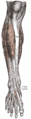 Gray's Anatomy illustration of the inferior extensor retinaculum of the foot
 -
 Slide showing the inferior extensor retinaculum of the foot


Medical Disclaimer: WikiMD is for informational purposes only and is not a substitute for professional medical advice. Content may be inaccurate or outdated and should not be used for diagnosis or treatment. Always consult your healthcare provider for medical decisions. Verify information with trusted sources such as CDC.gov and NIH.gov. By using this site, you agree that WikiMD is not liable for any outcomes related to its content. See full disclaimer.
Credits:Most images are courtesy of Wikimedia commons, and templates, categories Wikipedia, licensed under CC BY SA or similar.
Translate page: - East Asian
中文,
日本,
한국어,
South Asian
हिन्दी,
தமிழ்,
తెలుగు,
Urdu,
ಕನ್ನಡ,
Southeast Asian
Indonesian,
Vietnamese,
Thai,
မြန်မာဘာသာ,
বাংলা
European
español,
Deutsch,
français,
Greek,
português do Brasil,
polski,
română,
русский,
Nederlands,
norsk,
svenska,
suomi,
Italian
Middle Eastern & African
عربى,
Turkish,
Persian,
Hebrew,
Afrikaans,
isiZulu,
Kiswahili,
Other
Bulgarian,
Hungarian,
Czech,
Swedish,
മലയാളം,
मराठी,
ਪੰਜਾਬੀ,
ગુજરાતી,
Portuguese,
Ukrainian
