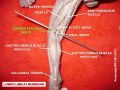
Common fibular nerve
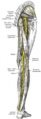
The common fibular nerve, also known as the peroneal nerve, is a major nerve in the lower limb. It is one of the two terminal branches of the sciatic nerve, along with the tibial nerve. The common fibular nerve innervates several muscles in the leg and provides sensory innervation to the skin of the lower leg and foot.
Anatomy[edit]
The common fibular nerve arises from the sciatic nerve in the posterior thigh. It originates from the lumbosacral plexus, specifically from the ventral rami of the fourth and fifth lumbar nerves (L4-L5) and the first and second sacral nerves (S1-S2). It descends along the posterior aspect of the thigh, passing through the popliteal fossa.
In the popliteal fossa, the common fibular nerve divides into two branches: the superficial fibular nerve and the deep fibular nerve. The superficial fibular nerve continues its course laterally, while the deep fibular nerve descends anteriorly.
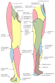
The superficial fibular nerve innervates the muscles in the lateral compartment of the leg, including the fibularis longus and fibularis brevis muscles. It also provides sensory innervation to the skin on the dorsum of the foot and the lateral aspect of the lower leg.
The deep fibular nerve innervates the muscles in the anterior compartment of the leg, including the tibialis anterior, extensor hallucis longus, extensor digitorum longus, and fibularis tertius muscles. It also provides sensory innervation to the skin between the first and second toes.
Clinical Significance[edit]
Injury or compression of the common fibular nerve can lead to various symptoms and conditions. Common causes of common fibular nerve injury include trauma, compression, and nerve entrapment.
One common condition associated with common fibular nerve injury is foot drop. Foot drop refers to the inability to dorsiflex the foot and toes, resulting in a characteristic gait abnormality. This can occur due to damage to the deep fibular nerve, which innervates the muscles responsible for dorsiflexion.
Another condition associated with common fibular nerve injury is peroneal neuropathy. Peroneal neuropathy is characterized by weakness or paralysis of the muscles in the lateral compartment of the leg, as well as sensory loss in the affected area.
Treatment[edit]
Treatment for common fibular nerve injury depends on the underlying cause and severity of the condition. Conservative management may include rest, physical therapy, and the use of assistive devices such as braces or splints to support the affected limb.
In more severe cases, surgical intervention may be necessary to decompress or repair the damaged nerve. This can help restore function and alleviate symptoms associated with common fibular nerve injury.
See Also[edit]
References[edit]
Common_fibular_nerve[edit]
-
 Common fibular nerve
 -
 Common fibular nerve
 -
Common fibular nerve
-
 Common fibular nerve
 -
Common fibular nerve


Medical Disclaimer: WikiMD is for informational purposes only and is not a substitute for professional medical advice. Content may be inaccurate or outdated and should not be used for diagnosis or treatment. Always consult your healthcare provider for medical decisions. Verify information with trusted sources such as CDC.gov and NIH.gov. By using this site, you agree that WikiMD is not liable for any outcomes related to its content. See full disclaimer.
Credits:Most images are courtesy of Wikimedia commons, and templates, categories Wikipedia, licensed under CC BY SA or similar.
Translate page: - East Asian
中文,
日本,
한국어,
South Asian
हिन्दी,
தமிழ்,
తెలుగు,
Urdu,
ಕನ್ನಡ,
Southeast Asian
Indonesian,
Vietnamese,
Thai,
မြန်မာဘာသာ,
বাংলা
European
español,
Deutsch,
français,
Greek,
português do Brasil,
polski,
română,
русский,
Nederlands,
norsk,
svenska,
suomi,
Italian
Middle Eastern & African
عربى,
Turkish,
Persian,
Hebrew,
Afrikaans,
isiZulu,
Kiswahili,
Other
Bulgarian,
Hungarian,
Czech,
Swedish,
മലയാളം,
मराठी,
ਪੰਜਾਬੀ,
ગુજરાતી,
Portuguese,
Ukrainian
