Amniotic sac
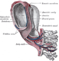
The amniotic sac, also known as the bag of waters or the membranes, is a thin but tough transparent pair of membranes that hold a developing embryo (and later fetus) until shortly before birth. The inner of these fetal membranes, the amnion, encloses the amniotic fluid and the fetus. The outer membrane, the chorion, contains the amnion and is part of the placenta. On the outer side, the amniotic sac is connected to the yolk sac, the allantois, and, via the umbilical cord, to the placenta.
Structure[edit]
The amniotic sac is composed of two membranes, the amnion and the chorion. The amnion, the inner membrane, is in contact with the amniotic fluid and the fetus. The chorion, the outer membrane, is in contact with maternal tissues and contributes to the formation of the placenta.
Function[edit]
The primary role of the amniotic sac is to protect the developing fetus by cushioning against physical trauma, providing a stable temperature, and preventing desiccation. It also allows the fetus to move freely within the womb, promoting muscular and skeletal development.
Clinical significance[edit]
The amniotic sac is clinically significant in several ways. It is involved in many pregnancy complications, such as preterm premature rupture of membranes (PPROM), where the amniotic sac breaks before 37 weeks of gestation, and oligohydramnios, a condition characterized by too little amniotic fluid.
See also[edit]
This medical article is a stub. You can help WikiMD by expanding the page. |
-
 Amniotic sac
 -
Bilaminar embryonic disc at 14 days in the implantation site in endometrium
-
 Amnion
 -
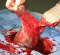
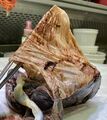 Placenta with fetal membranes


Medical Disclaimer: WikiMD is for informational purposes only and is not a substitute for professional medical advice. Content may be inaccurate or outdated and should not be used for diagnosis or treatment. Always consult your healthcare provider for medical decisions. Verify information with trusted sources such as CDC.gov and NIH.gov. By using this site, you agree that WikiMD is not liable for any outcomes related to its content. See full disclaimer.
Credits:Most images are courtesy of Wikimedia commons, and templates, categories Wikipedia, licensed under CC BY SA or similar.
Translate page: - East Asian
中文,
日本,
한국어,
South Asian
हिन्दी,
தமிழ்,
తెలుగు,
Urdu,
ಕನ್ನಡ,
Southeast Asian
Indonesian,
Vietnamese,
Thai,
မြန်မာဘာသာ,
বাংলা
European
español,
Deutsch,
français,
Greek,
português do Brasil,
polski,
română,
русский,
Nederlands,
norsk,
svenska,
suomi,
Italian
Middle Eastern & African
عربى,
Turkish,
Persian,
Hebrew,
Afrikaans,
isiZulu,
Kiswahili,
Other
Bulgarian,
Hungarian,
Czech,
Swedish,
മലയാളം,
मराठी,
ਪੰਜਾਬੀ,
ગુજરાતી,
Portuguese,
Ukrainian
