Atomic force microscopy


Atomic Force Microscopy (AFM) is a type of scanning probe microscopy (SPM) that provides a 3D profile of a surface on a nanoscale. The resolution of AFM is in the order of fractions of a nanometer, more than 1000 times better than the optical diffraction limit. AFM is a key tool in various fields including materials science, biophysics, and nanotechnology, allowing for the imaging, measuring, and manipulation of surfaces at the atomic scale.
History[edit]
AFM was invented in 1986 by Gerd Binnig, Calvin Quate, and Christoph Gerber. This invention was a breakthrough in the field of microscopy as it allowed scientists to visualize surfaces at an atomic level without the need for vacuum environments, unlike electron microscopes.
Principle[edit]
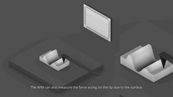
The basic working principle of AFM involves a cantilever with a sharp tip (probe) at its end that is used to scan the specimen surface. The cantilever is typically made of silicon or silicon nitride with a tip radius of curvature on the order of nanometers. When the tip is brought into proximity of a sample surface, forces between the tip and the surface lead to a deflection of the cantilever according to Hooke's law. These deflections are measured using a laser beam that is reflected off the top surface of the cantilever into an array of photodiodes.
Modes of Operation[edit]
AFM can operate in several modes, depending on the application:
- Contact mode involves the tip being in constant contact with the sample surface, used for measuring physical properties like hardness.
- Tapping mode (also known as intermittent contact mode) reduces the damage to the sample by only touching the surface at certain intervals.
- Non-contact mode measures the force between the tip and the sample without actual contact, useful for soft or sticky surfaces.
Applications[edit]
AFM has a wide range of applications across various scientific disciplines:
- In materials science, it is used to study the surface structure, properties, and defects of materials.
- In biology, AFM helps in imaging cells and tissues, and measuring mechanical properties of biological molecules.
- In nanotechnology, it is utilized for the manipulation of atoms and molecules to create nanostructures.
Advantages and Limitations[edit]
The main advantage of AFM is its ability to image non-conducting materials without any special preparation. However, its limitations include a relatively slow scanning speed and the potential for the tip to modify the sample surface during scanning.
Future Directions[edit]
Research in AFM technology is focused on improving the speed of scanning, enhancing resolution, and developing new modes for specific applications. Innovations such as high-speed AFM and multifrequency AFM techniques are expanding the capabilities and applications of atomic force microscopy.


Medical Disclaimer: WikiMD is for informational purposes only and is not a substitute for professional medical advice. Content may be inaccurate or outdated and should not be used for diagnosis or treatment. Always consult your healthcare provider for medical decisions. Verify information with trusted sources such as CDC.gov and NIH.gov. By using this site, you agree that WikiMD is not liable for any outcomes related to its content. See full disclaimer.
Credits:Most images are courtesy of Wikimedia commons, and templates, categories Wikipedia, licensed under CC BY SA or similar.
Translate page: - East Asian
中文,
日本,
한국어,
South Asian
हिन्दी,
தமிழ்,
తెలుగు,
Urdu,
ಕನ್ನಡ,
Southeast Asian
Indonesian,
Vietnamese,
Thai,
မြန်မာဘာသာ,
বাংলা
European
español,
Deutsch,
français,
Greek,
português do Brasil,
polski,
română,
русский,
Nederlands,
norsk,
svenska,
suomi,
Italian
Middle Eastern & African
عربى,
Turkish,
Persian,
Hebrew,
Afrikaans,
isiZulu,
Kiswahili,
Other
Bulgarian,
Hungarian,
Czech,
Swedish,
മലയാളം,
मराठी,
ਪੰਜਾਬੀ,
ગુજરાતી,
Portuguese,
Ukrainian
