Bright-field microscopy
Bright-field microscopy is a fundamental microscopy technique used in biology, medicine, and various scientific fields to observe specimens with visible light. This method of microscopy is one of the simplest and most widely used techniques for examining the microstructure of specimens. Bright-field microscopy produces an image with a light background and darker sample features, making it ideal for observing stained or naturally pigmented samples.
Principles of Operation[edit]
Bright-field microscopy operates on the principle of transmitting light through a specimen. Light from an illumination source passes through a series of optical lenses, including the objective lens and the eyepiece, to magnify the image of the specimen. The key components of a bright-field microscope include the light source, condenser, specimen stage, objective lenses, and eyepiece or camera.
The condenser lens focuses the light onto the specimen, ensuring that the light rays are parallel as they pass through the sample. This illumination setup is critical for achieving optimal resolution and contrast in the observed image. The objective lens collects the light transmitted through the specimen and forms an enlarged image, which is further magnified by the eyepiece.
Sample Preparation[edit]
For effective observation under a bright-field microscope, specimens often require preparation to enhance contrast, as the technique inherently has low contrast for unstained or transparent samples. Common preparation methods include:
- Staining: Applying chemical dyes to the specimen to add color and enhance contrast between different components of the specimen.
- Fixation: Treating the specimen with chemicals to preserve its structure and prevent degradation.
- Sectioning: Cutting the specimen into thin slices suitable for light transmission.
Applications[edit]
Bright-field microscopy is widely used in various fields for its simplicity and effectiveness in observing stained specimens. Applications include:
- Biology: Observing cellular structures, bacteria, and tissue samples.
- Medicine: Diagnosing diseases by examining blood smears, biopsy samples, and other clinical specimens.
- Material Science: Analyzing the microstructure of materials, including metals, ceramics, and polymers.
Limitations[edit]
While bright-field microscopy is versatile, it has limitations, including:
- Low contrast for unstained or transparent specimens.
- Limited resolution due to the diffraction limit of light.
- Potential for photodamage to light-sensitive specimens.
Enhancements[edit]
Technological advancements have led to enhancements in bright-field microscopy, including:
- Phase contrast microscopy: A technique that converts phase shifts in light passing through a transparent specimen into changes in intensity, improving contrast without staining.
- Digital microscopy: Incorporating digital cameras and software for image capture, analysis, and sharing.
Conclusion[edit]
Bright-field microscopy remains a cornerstone technique in microscopy due to its simplicity, effectiveness, and wide range of applications. Despite its limitations, ongoing advancements continue to expand its capabilities and applications in scientific research and medical diagnostics.
This Microscopy-related article is a stub. You can help WikiMD by expanding the page. |
Bright-field microscopy[edit]
-
 Cross-section of a Zea stem at 100x magnification
 -
 Illustration of bees by Francesco Stelluti, 1630
 -
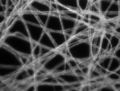
 Bright-field micrograph of paper fibers
 -
 Cross-polarised micrograph of paper fibers
 -
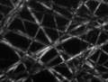
 Dark-field micrograph of paper fibers
 -
 Phase-contrast micrograph of paper fibers


Medical Disclaimer: WikiMD is for informational purposes only and is not a substitute for professional medical advice. Content may be inaccurate or outdated and should not be used for diagnosis or treatment. Always consult your healthcare provider for medical decisions. Verify information with trusted sources such as CDC.gov and NIH.gov. By using this site, you agree that WikiMD is not liable for any outcomes related to its content. See full disclaimer.
Credits:Most images are courtesy of Wikimedia commons, and templates, categories Wikipedia, licensed under CC BY SA or similar.
Translate page: - East Asian
中文,
日本,
한국어,
South Asian
हिन्दी,
தமிழ்,
తెలుగు,
Urdu,
ಕನ್ನಡ,
Southeast Asian
Indonesian,
Vietnamese,
Thai,
မြန်မာဘာသာ,
বাংলা
European
español,
Deutsch,
français,
Greek,
português do Brasil,
polski,
română,
русский,
Nederlands,
norsk,
svenska,
suomi,
Italian
Middle Eastern & African
عربى,
Turkish,
Persian,
Hebrew,
Afrikaans,
isiZulu,
Kiswahili,
Other
Bulgarian,
Hungarian,
Czech,
Swedish,
മലയാളം,
मराठी,
ਪੰਜਾਬੀ,
ગુજરાતી,
Portuguese,
Ukrainian
