Chorionic villi
Chorionic villi are microscopic, finger-like projections that emerge from the embryo's chorion to provide maximum contact area with the maternal blood. They are a critical part of the placenta, facilitating the exchange of nutrients, gases, and wastes between the mother and the fetus.
Structure[edit]
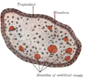
The chorionic villi are composed of a core of mesoderm cells, which includes blood vessels, surrounded by two layers of trophoblast cells. The outer layer, known as the syncytiotrophoblast, is in direct contact with maternal blood, while the inner layer, the cytotrophoblast, provides support.
Function[edit]
The primary function of the chorionic villi is to facilitate the exchange of nutrients, gases, and wastes between the mother and the fetus. The villi are bathed in maternal blood, and the thin walls of the villi allow for the easy diffusion of substances between the maternal and fetal bloodstreams.
In addition to this, the chorionic villi also produce several hormones, including human chorionic gonadotropin (hCG), which is crucial for maintaining pregnancy.
Clinical significance[edit]
Abnormalities in the development or function of the chorionic villi can lead to complications in pregnancy. For example, in placenta previa, the placenta implants too low in the uterus, potentially blocking the cervix and leading to bleeding.
Chorionic villus sampling (CVS) is a prenatal test where a small sample of chorionic villi is removed from the placenta for genetic testing. This can provide information about the baby's genetic health early in pregnancy.
See also[edit]

Chorionic_villi[edit]
-
Chorionic villus
-
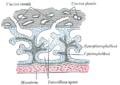 Gray's Anatomy illustration 36
 -
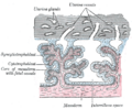 Gray's Anatomy illustration 37
 -
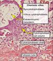 Histopathology of tubal pregnancy
 -
Gross pathology of fixed chorionic villi
-
Chorionic villi - intermediate magnification
-
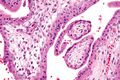 Chorionic villi - very high magnification
 -
 Gray's Anatomy illustration 21
 -
 Gray's Anatomy illustration 35
 -
 Gray's Anatomy illustration 459


Medical Disclaimer: WikiMD is for informational purposes only and is not a substitute for professional medical advice. Content may be inaccurate or outdated and should not be used for diagnosis or treatment. Always consult your healthcare provider for medical decisions. Verify information with trusted sources such as CDC.gov and NIH.gov. By using this site, you agree that WikiMD is not liable for any outcomes related to its content. See full disclaimer.
Credits:Most images are courtesy of Wikimedia commons, and templates, categories Wikipedia, licensed under CC BY SA or similar.
Translate page: - East Asian
中文,
日本,
한국어,
South Asian
हिन्दी,
தமிழ்,
తెలుగు,
Urdu,
ಕನ್ನಡ,
Southeast Asian
Indonesian,
Vietnamese,
Thai,
မြန်မာဘာသာ,
বাংলা
European
español,
Deutsch,
français,
Greek,
português do Brasil,
polski,
română,
русский,
Nederlands,
norsk,
svenska,
suomi,
Italian
Middle Eastern & African
عربى,
Turkish,
Persian,
Hebrew,
Afrikaans,
isiZulu,
Kiswahili,
Other
Bulgarian,
Hungarian,
Czech,
Swedish,
മലയാളം,
मराठी,
ਪੰਜਾਬੀ,
ગુજરાતી,
Portuguese,
Ukrainian
