Spinal nerve
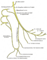
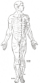
Spinal nerve is a mixed nerve, which carries motor, sensory, and autonomic signals between the spinal cord and the body. In the human body, there are 31 pairs of spinal nerves, one on each side of the vertebral column. These are grouped into the corresponding cervical, thoracic, lumbar, sacral and coccygeal regions of the spine. There are eight pairs of cervical nerves, twelve pairs of thoracic nerves, five pairs of lumbar nerves, five pairs of sacral nerves, and one pair of coccygeal nerves. The spinal nerves are part of the peripheral nervous system (PNS).
Structure[edit]
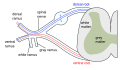
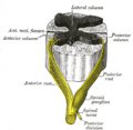
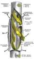
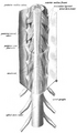
Each spinal nerve is a mixed nerve that is formed from the combination of nerve fibers from its posterior and anterior roots. The posterior root is the sensory root that carries sensory information to the brain from other parts of the body. The anterior root is the motor root that transmits neural signals from the brain to the various parts of the body.
Function[edit]
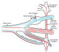
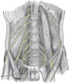
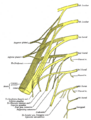
The spinal nerves provide the connection between the central nervous system and the nerves supplying the peripheral tissues. Each spinal nerve carries both sensory and motor information.
Clinical significance[edit]
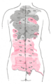
Damage to the spinal nerves can cause neurological disorders, such as radiculopathy, which is characterized by pain that radiates along the nerve from the spine. Other conditions, such as spinal cord injury, can also affect the spinal nerves.
See also[edit]

This WikiMD article can only be edited by registered and verified editors. You can log in or register.
-
 Spinal nerve
 -
 Spinal nerve
 -
 Spinal nerve
 -
 Spinal nerve
 -
 Spinal nerve
 -
 Spinal nerve
 -
 Spinal nerve
 -
 Spinal nerve
 -
 Spinal nerve
 -
 Spinal nerve
 -
 Spinal nerve
 -
 Spinal nerve


Medical Disclaimer: WikiMD is for informational purposes only and is not a substitute for professional medical advice. Content may be inaccurate or outdated and should not be used for diagnosis or treatment. Always consult your healthcare provider for medical decisions. Verify information with trusted sources such as CDC.gov and NIH.gov. By using this site, you agree that WikiMD is not liable for any outcomes related to its content. See full disclaimer.
Credits:Most images are courtesy of Wikimedia commons, and templates, categories Wikipedia, licensed under CC BY SA or similar.
Translate page: - East Asian
中文,
日本,
한국어,
South Asian
हिन्दी,
தமிழ்,
తెలుగు,
Urdu,
ಕನ್ನಡ,
Southeast Asian
Indonesian,
Vietnamese,
Thai,
မြန်မာဘာသာ,
বাংলা
European
español,
Deutsch,
français,
Greek,
português do Brasil,
polski,
română,
русский,
Nederlands,
norsk,
svenska,
suomi,
Italian
Middle Eastern & African
عربى,
Turkish,
Persian,
Hebrew,
Afrikaans,
isiZulu,
Kiswahili,
Other
Bulgarian,
Hungarian,
Czech,
Swedish,
മലയാളം,
मराठी,
ਪੰਜਾਬੀ,
ગુજરાતી,
Portuguese,
Ukrainian
