Cuneiform cartilages
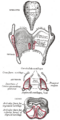
Cuneiform cartilages are small, paired cartilages located in the larynx, specifically within the aryepiglottic fold. They are part of the laryngeal skeleton, which is made up of nine cartilages. The cuneiform cartilages are among the three paired cartilages in the larynx, the other two being the arytenoid cartilages and the corniculate cartilages.
Etymology[edit]
The term "cuneiform" is derived from the Latin word "cuneus", which means "wedge". This is due to the wedge-like shape of these cartilages.
Structure[edit]
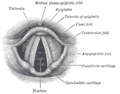
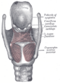
The cuneiform cartilages are small, elongated pieces of yellow elastic cartilage. They are located in the aryepiglottic fold, which is a fold of mucous membrane running from the epiglottis to the arytenoid cartilages. The cuneiform cartilages provide support and stiffness to the aryepiglottic folds.
Function[edit]
The primary function of the cuneiform cartilages is to support the vocal apparatus and to help maintain the opening of the larynx. They also contribute to the structure of the epiglottic vallecula, a space at the base of the tongue between the folds of the epiglottis.
Clinical significance[edit]
Abnormalities or injuries to the cuneiform cartilages can affect voice production and breathing. For example, laryngeal cancer can involve the cuneiform cartilages, leading to symptoms such as hoarseness, difficulty swallowing, and shortness of breath.
See also[edit]

This WikiMD article can only be edited by registered and verified editors. You can log in or register.
Cuneiform_cartilages[edit]
-
 Gray956
 -
 Gray950
 -
 Gray958


Medical Disclaimer: WikiMD is for informational purposes only and is not a substitute for professional medical advice. Content may be inaccurate or outdated and should not be used for diagnosis or treatment. Always consult your healthcare provider for medical decisions. Verify information with trusted sources such as CDC.gov and NIH.gov. By using this site, you agree that WikiMD is not liable for any outcomes related to its content. See full disclaimer.
Credits:Most images are courtesy of Wikimedia commons, and templates, categories Wikipedia, licensed under CC BY SA or similar.
Translate page: - East Asian
中文,
日本,
한국어,
South Asian
हिन्दी,
தமிழ்,
తెలుగు,
Urdu,
ಕನ್ನಡ,
Southeast Asian
Indonesian,
Vietnamese,
Thai,
မြန်မာဘာသာ,
বাংলা
European
español,
Deutsch,
français,
Greek,
português do Brasil,
polski,
română,
русский,
Nederlands,
norsk,
svenska,
suomi,
Italian
Middle Eastern & African
عربى,
Turkish,
Persian,
Hebrew,
Afrikaans,
isiZulu,
Kiswahili,
Other
Bulgarian,
Hungarian,
Czech,
Swedish,
മലയാളം,
मराठी,
ਪੰਜਾਬੀ,
ગુજરાતી,
Portuguese,
Ukrainian
