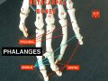
Phalanx bone
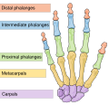
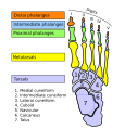
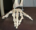
Phalanx bones' are the bones that form the fingers of the hands and the toes of the feet. Each finger and toe, except for the thumb and big toe, has three phalanx bones, which are referred to as the proximal, middle, and distal phalanges. The thumb and big toe have only two phalanx bones, lacking the middle phalanx. These bones play a crucial role in the movement and dexterity of the hands and feet.
Structure[edit]
The structure of the phalanx bones is similar to most long bones in the body, having a shaft and two ends. The ends are known as the base and the head. The base is the part of the bone that is closer to the wrist or ankle, and the head is the part that is closer to the fingertips or toes. The surface of the phalanx bones is covered with articular cartilage at the joints, which helps in smooth movement and reduces friction.
Function[edit]
The primary function of the phalanx bones is to provide support and flexibility to the fingers and toes. These bones work in conjunction with the muscles and tendons in the hand and foot to allow for a wide range of movements, from grasping objects to walking. The arrangement of the phalanges also contributes to the gripping ability and the fine motor skills of the hands.
Clinical Significance[edit]
Phalanx bones are prone to fractures, which are common injuries that can occur from direct impacts or falls. Treatment for phalanx fractures depends on the severity and location of the fracture and may include immobilization, physical therapy, or surgery. Conditions such as osteoarthritis can also affect the phalanx bones, leading to pain and reduced mobility.
Development[edit]
The development of the phalanx bones begins in the embryonic stage with the process of endochondral ossification. This is where cartilage is gradually replaced by bone. The growth of the phalanges continues throughout childhood and adolescence, with growth plates located at the ends of the bones.
Evolution[edit]
The phalanx bones have evolved to support the diverse range of functions of the hands and feet in different species. In humans, the structure of the phalanges allows for precise movements and manipulation of objects, which is a key aspect of human tool use and cultural development.
This medical article is a stub. You can help WikiMD by expanding the page. |
-
 Phalanx_bone
 -
 Phalanx_bone
 -
 Phalanx_bone
 -
 Phalanx_bone
 -
 Phalanx_bone
 -
 Phalanx_bone
 -
 Phalanx_bone
 -
 Phalanx_bone
 -
 Phalanx_bone
 -
 Phalanx_bone
 -
 Phalanx_bone
 -
 Phalanx_bone


Medical Disclaimer: WikiMD is for informational purposes only and is not a substitute for professional medical advice. Content may be inaccurate or outdated and should not be used for diagnosis or treatment. Always consult your healthcare provider for medical decisions. Verify information with trusted sources such as CDC.gov and NIH.gov. By using this site, you agree that WikiMD is not liable for any outcomes related to its content. See full disclaimer.
Credits:Most images are courtesy of Wikimedia commons, and templates, categories Wikipedia, licensed under CC BY SA or similar.
Translate page: - East Asian
中文,
日本,
한국어,
South Asian
हिन्दी,
தமிழ்,
తెలుగు,
Urdu,
ಕನ್ನಡ,
Southeast Asian
Indonesian,
Vietnamese,
Thai,
မြန်မာဘာသာ,
বাংলা
European
español,
Deutsch,
français,
Greek,
português do Brasil,
polski,
română,
русский,
Nederlands,
norsk,
svenska,
suomi,
Italian
Middle Eastern & African
عربى,
Turkish,
Persian,
Hebrew,
Afrikaans,
isiZulu,
Kiswahili,
Other
Bulgarian,
Hungarian,
Czech,
Swedish,
മലയാളം,
मराठी,
ਪੰਜਾਬੀ,
ગુજરાતી,
Portuguese,
Ukrainian
