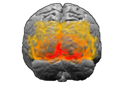
Extrastriate cortex
Extrastriate Cortex[edit | edit source]
The extrastriate cortex is a region of the cerebral cortex located in the occipital lobe of the brain, adjacent to the primary visual cortex (V1). It is involved in the processing of visual information and is crucial for the perception of complex visual stimuli. The extrastriate cortex includes several distinct areas, each with specialized functions in visual processing.
Anatomy[edit | edit source]
The extrastriate cortex encompasses several Brodmann areas, primarily areas 18 and 19, which are located around the primary visual cortex (Brodmann area 17). These areas are part of the visual cortex and are involved in higher-order visual processing. The extrastriate cortex is divided into multiple regions, each responsible for different aspects of visual perception, such as motion, color, and form.
Brodmann Area 18[edit | edit source]
Brodmann area 18, also known as the secondary visual cortex or V2, is located adjacent to the primary visual cortex. It receives input from V1 and is involved in the processing of visual information related to orientation, spatial frequency, and color. V2 is organized into stripes, each of which processes different types of visual information.
Brodmann Area 19[edit | edit source]
Brodmann area 19, also known as the tertiary visual cortex or V3, is involved in the processing of complex visual stimuli, such as motion and depth perception. It receives input from both V1 and V2 and is connected to other visual areas in the brain, including the dorsal stream and ventral stream.
Function[edit | edit source]
The extrastriate cortex plays a critical role in the interpretation and integration of visual information. It is involved in the perception of motion, depth, color, and form. The extrastriate cortex is also important for visual attention and the recognition of objects and faces.
Visual Processing Streams[edit | edit source]
The extrastriate cortex is part of two major visual processing streams:
- The dorsal stream, also known as the "where" pathway, is involved in the processing of spatial location and motion. It extends from the extrastriate cortex to the parietal lobe.
- The ventral stream, also known as the "what" pathway, is involved in the recognition and identification of objects. It extends from the extrastriate cortex to the temporal lobe.
Clinical Significance[edit | edit source]
Damage to the extrastriate cortex can result in various visual disorders, depending on the specific area affected. For example, lesions in V2 can lead to deficits in color perception, while damage to V3 can impair motion perception. Disorders such as visual agnosia and prosopagnosia are associated with dysfunction in the extrastriate cortex.
Related Pages[edit | edit source]
Search WikiMD
Ad.Tired of being Overweight? Try W8MD's physician weight loss program.
Semaglutide (Ozempic / Wegovy and Tirzepatide (Mounjaro / Zepbound) available.
Advertise on WikiMD
|
WikiMD's Wellness Encyclopedia |
| Let Food Be Thy Medicine Medicine Thy Food - Hippocrates |
Translate this page: - East Asian
中文,
日本,
한국어,
South Asian
हिन्दी,
தமிழ்,
తెలుగు,
Urdu,
ಕನ್ನಡ,
Southeast Asian
Indonesian,
Vietnamese,
Thai,
မြန်မာဘာသာ,
বাংলা
European
español,
Deutsch,
français,
Greek,
português do Brasil,
polski,
română,
русский,
Nederlands,
norsk,
svenska,
suomi,
Italian
Middle Eastern & African
عربى,
Turkish,
Persian,
Hebrew,
Afrikaans,
isiZulu,
Kiswahili,
Other
Bulgarian,
Hungarian,
Czech,
Swedish,
മലയാളം,
मराठी,
ਪੰਜਾਬੀ,
ગુજરાતી,
Portuguese,
Ukrainian
Medical Disclaimer: WikiMD is not a substitute for professional medical advice. The information on WikiMD is provided as an information resource only, may be incorrect, outdated or misleading, and is not to be used or relied on for any diagnostic or treatment purposes. Please consult your health care provider before making any healthcare decisions or for guidance about a specific medical condition. WikiMD expressly disclaims responsibility, and shall have no liability, for any damages, loss, injury, or liability whatsoever suffered as a result of your reliance on the information contained in this site. By visiting this site you agree to the foregoing terms and conditions, which may from time to time be changed or supplemented by WikiMD. If you do not agree to the foregoing terms and conditions, you should not enter or use this site. See full disclaimer.
Credits:Most images are courtesy of Wikimedia commons, and templates, categories Wikipedia, licensed under CC BY SA or similar.
Contributors: Prab R. Tumpati, MD

