H&E stain
H&E stain or Hematoxylin and Eosin stain is a popular staining method in histology. It is named after the dyes hematoxylin and eosin, which it uses. Hematoxylin, which stains nuclei blue-purple, and eosin, which stains cytoplasm and the extracellular connective tissue matrix pink, are used.
Overview[edit]
H&E staining is used to examine tissues under a microscope and is one of the most commonly used stains in medical diagnosis. For example, it is often used to diagnose cancer. A pathologist will look at the tissue sample under a microscope and make a diagnosis based on the size, shape, and organization of the cells.
Procedure[edit]
The procedure for H&E staining is as follows:
- The tissue is fixed in a substance such as formalin to preserve it.
- The tissue is then cut into thin slices, or sections. These sections are placed on a glass slide.
- The slide is then stained with hematoxylin, which stains the cell nuclei blue.
- The slide is then stained with eosin, which stains the cytoplasm and connective tissue pink.
- The slide is then examined under a microscope.
Interpretation[edit]
The interpretation of H&E staining involves looking at the size, shape, and organization of the cells. Normal cells are uniform in size and shape, and are organized in a predictable pattern. Cancer cells, on the other hand, are often larger than normal cells, are irregular in shape, and are not organized in a predictable pattern.
See also[edit]

This WikiMD article can only be edited by registered and verified editors. You can log in or register.
-
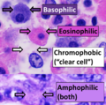 Eosinophilic, basophilic, chromophobic and amphophilic staining
 -
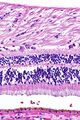 Retina -- high magnification
 -
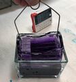 Slide rack in haematoxylin
 -
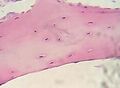 Cartilage
 -
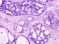 Breast DCIS histopathology
 -
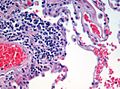 Emphysema H and E
 -
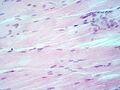 Musc est long 400
 -
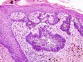 Basal cell carcinoma histopathology


.jpg)


.jpg)

Medical Disclaimer: WikiMD is for informational purposes only and is not a substitute for professional medical advice. Content may be inaccurate or outdated and should not be used for diagnosis or treatment. Always consult your healthcare provider for medical decisions. Verify information with trusted sources such as CDC.gov and NIH.gov. By using this site, you agree that WikiMD is not liable for any outcomes related to its content. See full disclaimer.
Credits:Most images are courtesy of Wikimedia commons, and templates, categories Wikipedia, licensed under CC BY SA or similar.
Translate page: - East Asian
中文,
日本,
한국어,
South Asian
हिन्दी,
தமிழ்,
తెలుగు,
Urdu,
ಕನ್ನಡ,
Southeast Asian
Indonesian,
Vietnamese,
Thai,
မြန်မာဘာသာ,
বাংলা
European
español,
Deutsch,
français,
Greek,
português do Brasil,
polski,
română,
русский,
Nederlands,
norsk,
svenska,
suomi,
Italian
Middle Eastern & African
عربى,
Turkish,
Persian,
Hebrew,
Afrikaans,
isiZulu,
Kiswahili,
Other
Bulgarian,
Hungarian,
Czech,
Swedish,
മലയാളം,
मराठी,
ਪੰਜਾਬੀ,
ગુજરાતી,
Portuguese,
Ukrainian
