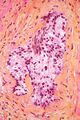
HPS stain
Hematoxylin Phloxine Saffron (HPS) Stain is a histological staining technique widely used in pathology to differentiate among various tissue types and to highlight specific structures within tissues. This staining method is particularly useful in the examination of biopsy specimens and in the diagnosis of various diseases, including cancer. The HPS stain is a modification of the traditional Hematoxylin and Eosin (H&E) stain, with the addition of saffron as a third component. This article provides a comprehensive overview of the HPS stain, including its components, application, and significance in medical diagnostics.
Components[edit]
The HPS stain consists of three main components, each serving a unique purpose in the staining process:
1. Hematoxylin: A basic dye that stains acidic, or basophilic, structures a blue or dark purple color. This includes cell nuclei and ribosomes, among other structures. 2. Phloxine: A synthetic dye that stains eosinophilic structures a bright pink color. This includes cytoplasm, keratin, and muscle fibers. 3. Saffron: A natural dye extracted from the crocus flower, used to stain collagen and connective tissues a yellow color.
Application[edit]
The application of HPS stain follows a specific protocol, which includes the following steps:
1. Fixation: Tissue samples are fixed, usually in formalin, to preserve their structure. 2. Embedding: The fixed tissue is embedded in paraffin wax to facilitate thin sectioning. 3. Sectioning: Thin sections (usually 4-5 micrometers thick) are cut from the paraffin-embedded tissue blocks. 4. Deparaffinization and Rehydration: Sections are treated with xylene or a similar solvent to remove paraffin, followed by a graded series of alcohol to rehydrate the tissues. 5. Staining: The tissue sections are stained with hematoxylin, followed by differentiation and bluing. Phloxine and saffron are then applied to stain other tissue components. 6. Dehydration and Mounting: Finally, the stained sections are dehydrated with alcohol, cleared in xylene, and mounted with a resinous medium for microscopic examination.
Significance[edit]
The HPS stain is invaluable in histopathology for several reasons:
- It provides excellent contrast between different tissue components, facilitating the identification of pathological changes. - The addition of saffron allows for better differentiation of connective tissues compared to the standard H&E stain. - It is particularly useful in the diagnosis of neoplastic diseases, as it highlights the architectural patterns of tumors and the relationship between tumor cells and the surrounding stroma.
Conclusion[edit]
The Hematoxylin Phloxine Saffron (HPS) Stain is a crucial tool in the field of pathology, offering detailed insights into tissue morphology and pathology. Its ability to differentiate among various tissue components with high contrast makes it an essential technique for the accurate diagnosis of diseases, particularly in the analysis of biopsy specimens.
-
 HPS stain


Medical Disclaimer: WikiMD is for informational purposes only and is not a substitute for professional medical advice. Content may be inaccurate or outdated and should not be used for diagnosis or treatment. Always consult your healthcare provider for medical decisions. Verify information with trusted sources such as CDC.gov and NIH.gov. By using this site, you agree that WikiMD is not liable for any outcomes related to its content. See full disclaimer.
Credits:Most images are courtesy of Wikimedia commons, and templates, categories Wikipedia, licensed under CC BY SA or similar.
Translate page: - East Asian
中文,
日本,
한국어,
South Asian
हिन्दी,
தமிழ்,
తెలుగు,
Urdu,
ಕನ್ನಡ,
Southeast Asian
Indonesian,
Vietnamese,
Thai,
မြန်မာဘာသာ,
বাংলা
European
español,
Deutsch,
français,
Greek,
português do Brasil,
polski,
română,
русский,
Nederlands,
norsk,
svenska,
suomi,
Italian
Middle Eastern & African
عربى,
Turkish,
Persian,
Hebrew,
Afrikaans,
isiZulu,
Kiswahili,
Other
Bulgarian,
Hungarian,
Czech,
Swedish,
മലയാളം,
मराठी,
ਪੰਜਾਬੀ,
ગુજરાતી,
Portuguese,
Ukrainian
