Iris dilator muscle


Muscle in the eye responsible for dilating the pupil
[[File: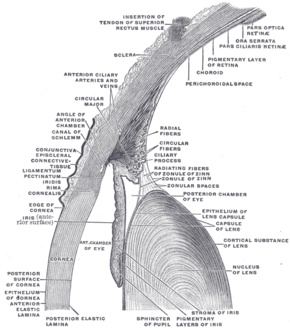 |frameless|alt=]]

| Details | |
|---|---|
| Synonyms | |
| Pronunciation | |
| Carnegie stage | |
| Days | |
| Precursor | |
| Gives rise to | |
| Part of | |
The iris dilator muscle (musculus dilatator pupillae) is a muscle located in the iris of the eye. It is responsible for dilating the pupil, allowing more light to enter the retina.
Structure[edit]
The iris dilator muscle is composed of a thin layer of myoepithelial cells that are radially arranged. These cells are located in the anterior part of the iris, just beneath the iris stroma. The muscle fibers extend from the pupillary margin to the periphery of the iris.
Innervation[edit]
The iris dilator muscle is innervated by the sympathetic nervous system. The preganglionic sympathetic fibers originate in the spinal cord at the level of T1 to T2 and travel to the superior cervical ganglion. From there, the postganglionic fibers travel along the internal carotid artery and enter the eye via the long ciliary nerves.
Function[edit]
The primary function of the iris dilator muscle is to dilate the pupil, a process known as mydriasis. This occurs in response to low light conditions or during the fight-or-flight response, allowing more light to reach the retina and improving vision in dim lighting.
Antagonist[edit]
The antagonist of the iris dilator muscle is the iris sphincter muscle, which constricts the pupil (a process known as miosis). The balance between these two muscles controls the size of the pupil.
Clinical significance[edit]
Dysfunction of the iris dilator muscle can lead to abnormal pupil responses. Conditions such as Horner's syndrome can result in a constricted pupil due to a lack of sympathetic innervation. Conversely, excessive stimulation of the sympathetic nervous system can cause prolonged pupil dilation.
See also[edit]


Medical Disclaimer: WikiMD is for informational purposes only and is not a substitute for professional medical advice. Content may be inaccurate or outdated and should not be used for diagnosis or treatment. Always consult your healthcare provider for medical decisions. Verify information with trusted sources such as CDC.gov and NIH.gov. By using this site, you agree that WikiMD is not liable for any outcomes related to its content. See full disclaimer.
Credits:Most images are courtesy of Wikimedia commons, and templates, categories Wikipedia, licensed under CC BY SA or similar.
Translate page: - East Asian
中文,
日本,
한국어,
South Asian
हिन्दी,
தமிழ்,
తెలుగు,
Urdu,
ಕನ್ನಡ,
Southeast Asian
Indonesian,
Vietnamese,
Thai,
မြန်မာဘာသာ,
বাংলা
European
español,
Deutsch,
français,
Greek,
português do Brasil,
polski,
română,
русский,
Nederlands,
norsk,
svenska,
suomi,
Italian
Middle Eastern & African
عربى,
Turkish,
Persian,
Hebrew,
Afrikaans,
isiZulu,
Kiswahili,
Other
Bulgarian,
Hungarian,
Czech,
Swedish,
മലയാളം,
मराठी,
ਪੰਜਾਬੀ,
ગુજરાતી,
Portuguese,
Ukrainian
