Superior rectus muscle
One of the extraocular muscles responsible for eye movement
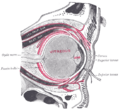
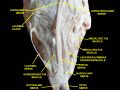
The superior rectus muscle is one of the six extraocular muscles that control the movements of the eye. It is primarily responsible for the elevation of the eye, allowing it to look upward. This muscle plays a crucial role in the complex coordination required for proper binocular vision.
Anatomy[edit]
The superior rectus muscle is located in the orbit, the bony cavity that houses the eye. It originates from the annulus of Zinn, a tendinous ring located at the apex of the orbit. From its origin, the muscle extends forward and slightly upward, inserting into the superior aspect of the sclera, the white outer coating of the eyeball, just posterior to the cornea.
The muscle is innervated by the oculomotor nerve (cranial nerve III), which provides the necessary motor signals for its contraction. The blood supply to the superior rectus muscle is primarily from the ophthalmic artery, a branch of the internal carotid artery.
Function[edit]
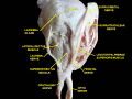
The primary function of the superior rectus muscle is to elevate the eye, moving the pupil upward. In addition to elevation, the muscle also contributes to intorsion (rotation of the top of the eye toward the nose) and adduction (movement of the eye toward the midline). These actions are essential for coordinated eye movements and maintaining proper alignment of the visual axes.
The superior rectus muscle works in conjunction with other extraocular muscles, such as the inferior rectus muscle, medial rectus muscle, and lateral rectus muscle, to achieve smooth and precise eye movements. The superior oblique muscle and inferior oblique muscle also assist in complex eye movements, particularly those involving torsion.
Clinical significance[edit]
Dysfunction of the superior rectus muscle can lead to various ocular motility disorders. For instance, strabismus, a condition characterized by misalignment of the eyes, can occur if the muscle is weak or paralyzed. This may result in diplopia (double vision) and difficulty with depth perception.
Injuries or lesions affecting the oculomotor nerve can impair the function of the superior rectus muscle, leading to a condition known as oculomotor nerve palsy. This condition can cause the affected eye to deviate downward and outward, as the unopposed action of the lateral rectus and superior oblique muscles take over.
Related pages[edit]
| Extraocular muscles | ||||||
|---|---|---|---|---|---|---|
This extraocular muscles related article is a stub.
|
Superior rectus muscle[edit]
-
 Gray891
 -
 Trochlear and frontal nerves
 -
Slide2uu
-
 Slide1abaa
 -
 Slide2abaa
 -
 Slide4abab
 -
 Slide6abab
 -
Slide8ababa


Medical Disclaimer: WikiMD is for informational purposes only and is not a substitute for professional medical advice. Content may be inaccurate or outdated and should not be used for diagnosis or treatment. Always consult your healthcare provider for medical decisions. Verify information with trusted sources such as CDC.gov and NIH.gov. By using this site, you agree that WikiMD is not liable for any outcomes related to its content. See full disclaimer.
Credits:Most images are courtesy of Wikimedia commons, and templates, categories Wikipedia, licensed under CC BY SA or similar.
Translate page: - East Asian
中文,
日本,
한국어,
South Asian
हिन्दी,
தமிழ்,
తెలుగు,
Urdu,
ಕನ್ನಡ,
Southeast Asian
Indonesian,
Vietnamese,
Thai,
မြန်မာဘာသာ,
বাংলা
European
español,
Deutsch,
français,
Greek,
português do Brasil,
polski,
română,
русский,
Nederlands,
norsk,
svenska,
suomi,
Italian
Middle Eastern & African
عربى,
Turkish,
Persian,
Hebrew,
Afrikaans,
isiZulu,
Kiswahili,
Other
Bulgarian,
Hungarian,
Czech,
Swedish,
മലയാളം,
मराठी,
ਪੰਜਾਬੀ,
ગુજરાતી,
Portuguese,
Ukrainian
