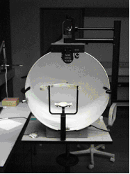
Visual field test
(Redirected from Visual field testing)
Visual field test
A visual field test is a method of measuring an individual's entire scope of vision, that is, their central vision and peripheral vision. This test is crucial in diagnosing and monitoring various eye diseases and neurological disorders.
Purpose[edit | edit source]
The primary purpose of a visual field test is to detect any abnormalities in the visual field. It is commonly used to diagnose and monitor conditions such as glaucoma, optic neuritis, stroke, and brain tumors. It can also help in assessing the visual function in patients with retinal diseases and optic nerve disorders.
Types of Visual Field Tests[edit | edit source]
There are several types of visual field tests, each with its own specific applications:
Confrontation Visual Field Test[edit | edit source]
The confrontation visual field test is a basic and quick screening test. The examiner compares the patient's visual field with their own by having the patient cover one eye and describe objects in their peripheral vision.
Automated Perimetry[edit | edit source]
Automated perimetry is a more advanced and precise method. It uses a machine to project light spots in different areas of the visual field. The patient presses a button each time they see a light spot. This test can map out the visual field in detail and is commonly used in diagnosing and monitoring glaucoma.
Kinetic Perimetry[edit | edit source]
Kinetic perimetry involves moving a stimulus from a non-seeing area to a seeing area. The patient indicates when they first see the stimulus. This test is useful for detecting visual field defects that may not be apparent in static tests.
Amsler Grid[edit | edit source]
The Amsler grid test is used primarily to detect macular degeneration and other central vision problems. The patient looks at a grid of horizontal and vertical lines and reports any distortions or missing areas.
Procedure[edit | edit source]
During a visual field test, the patient is usually seated and asked to focus on a central point. Depending on the type of test, they may be required to press a button or verbally indicate when they see a stimulus. The results are then analyzed to identify any areas of vision loss.
Interpretation of Results[edit | edit source]
The results of a visual field test are typically presented as a map of the visual field. Areas where the patient did not see the stimulus are marked as defects. These defects can indicate the presence of various eye or neurological conditions. For example, a pattern of vision loss in glaucoma often starts in the peripheral vision and progresses inward.
Clinical Significance[edit | edit source]
Visual field tests are essential tools in the early detection and management of eye diseases. Early detection of conditions like glaucoma can prevent significant vision loss. Regular visual field testing is recommended for individuals at risk of eye diseases, such as those with a family history of glaucoma or other risk factors.
See Also[edit | edit source]
References[edit | edit source]
External Links[edit | edit source]
Search WikiMD
Ad.Tired of being Overweight? Try W8MD's physician weight loss program.
Semaglutide (Ozempic / Wegovy and Tirzepatide (Mounjaro / Zepbound) available.
Advertise on WikiMD
|
WikiMD's Wellness Encyclopedia |
| Let Food Be Thy Medicine Medicine Thy Food - Hippocrates |
Translate this page: - East Asian
中文,
日本,
한국어,
South Asian
हिन्दी,
தமிழ்,
తెలుగు,
Urdu,
ಕನ್ನಡ,
Southeast Asian
Indonesian,
Vietnamese,
Thai,
မြန်မာဘာသာ,
বাংলা
European
español,
Deutsch,
français,
Greek,
português do Brasil,
polski,
română,
русский,
Nederlands,
norsk,
svenska,
suomi,
Italian
Middle Eastern & African
عربى,
Turkish,
Persian,
Hebrew,
Afrikaans,
isiZulu,
Kiswahili,
Other
Bulgarian,
Hungarian,
Czech,
Swedish,
മലയാളം,
मराठी,
ਪੰਜਾਬੀ,
ગુજરાતી,
Portuguese,
Ukrainian
Medical Disclaimer: WikiMD is not a substitute for professional medical advice. The information on WikiMD is provided as an information resource only, may be incorrect, outdated or misleading, and is not to be used or relied on for any diagnostic or treatment purposes. Please consult your health care provider before making any healthcare decisions or for guidance about a specific medical condition. WikiMD expressly disclaims responsibility, and shall have no liability, for any damages, loss, injury, or liability whatsoever suffered as a result of your reliance on the information contained in this site. By visiting this site you agree to the foregoing terms and conditions, which may from time to time be changed or supplemented by WikiMD. If you do not agree to the foregoing terms and conditions, you should not enter or use this site. See full disclaimer.
Credits:Most images are courtesy of Wikimedia commons, and templates, categories Wikipedia, licensed under CC BY SA or similar.
Contributors: Prab R. Tumpati, MD


