Anterior chamber of eyeball
Anterior Chamber of the Eyeball
The anterior chamber is a fluid-filled space inside the eye located between the cornea and the iris. It is one of the two chambers that make up the anterior segment of the eye, the other being the posterior chamber. The anterior chamber plays a crucial role in maintaining the eye's intraocular pressure through the production and drainage of aqueous humor, a clear fluid that nourishes the eye and maintains its shape.
Anatomy[edit]
The anterior chamber is bounded anteriorly by the cornea's innermost layer, the endothelium, and posteriorly by the iris and a portion of the lens's anterior capsule. Its depth can vary among individuals and changes with age, typically ranging from 2.5 to 3.5 mm in adults. The volume of the anterior chamber is approximately 250 µL.
Function[edit]
The primary function of the anterior chamber is to facilitate the circulation of aqueous humor, which is produced by the ciliary body located in the posterior chamber. Aqueous humor flows through the pupil from the posterior chamber into the anterior chamber and drains out of the eye via the trabecular meshwork and Schlemm's canal, helping to maintain a constant intraocular pressure. This fluid also provides nutrients to the avascular structures of the eye, such as the lens and cornea, and removes metabolic wastes.
Clinical Significance[edit]
Alterations in the anatomy or function of the anterior chamber can lead to various eye conditions. For example, a shallow anterior chamber may predispose an individual to angle-closure glaucoma, a condition where the outflow of aqueous humor is suddenly blocked, leading to a rapid increase in intraocular pressure. Conversely, a deep anterior chamber is often seen in cases of myopia or after certain types of eye surgery.
Hyphema, the accumulation of blood in the anterior chamber, is another condition that can occur due to trauma or as a complication of eye surgery. It can lead to increased intraocular pressure and vision loss if not treated promptly.
Diagnostic Techniques[edit]
Examination of the anterior chamber is an essential part of a comprehensive eye examination. Techniques such as slit lamp examination and gonioscopy are used to assess the depth of the anterior chamber, the angle of the anterior chamber (the angle formed between the iris and the cornea), and to detect any abnormalities. Ultrasound biomicroscopy (UBM) and anterior segment optical coherence tomography (AS-OCT) are advanced imaging techniques that provide detailed images of the anterior chamber and can help in diagnosing various conditions.
Treatment[edit]
Treatment of anterior chamber-related conditions depends on the underlying cause. For example, angle-closure glaucoma is treated with medications to lower intraocular pressure, laser therapy to improve the outflow of aqueous humor, or surgery to create a new drainage path. Hyphema is managed by resting the eye, elevating the head to promote drainage, and avoiding medications that can increase bleeding risk.
Conclusion[edit]
The anterior chamber of the eyeball is a vital structure involved in maintaining intraocular pressure and providing nutrients to the eye. Understanding its anatomy and function is essential for diagnosing and treating various eye conditions. Regular eye examinations are crucial for detecting changes in the anterior chamber that may indicate underlying eye diseases.
This medical article is a stub. You can help WikiMD by expanding the page. |
Anterior_chamber_of_eyeball[edit]
-
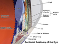 Cross-sectional anatomy of the eye
 -
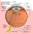 Schematic diagram of the human eye


Medical Disclaimer: WikiMD is for informational purposes only and is not a substitute for professional medical advice. Content may be inaccurate or outdated and should not be used for diagnosis or treatment. Always consult your healthcare provider for medical decisions. Verify information with trusted sources such as CDC.gov and NIH.gov. By using this site, you agree that WikiMD is not liable for any outcomes related to its content. See full disclaimer.
Credits:Most images are courtesy of Wikimedia commons, and templates, categories Wikipedia, licensed under CC BY SA or similar.
Translate page: - East Asian
中文,
日本,
한국어,
South Asian
हिन्दी,
தமிழ்,
తెలుగు,
Urdu,
ಕನ್ನಡ,
Southeast Asian
Indonesian,
Vietnamese,
Thai,
မြန်မာဘာသာ,
বাংলা
European
español,
Deutsch,
français,
Greek,
português do Brasil,
polski,
română,
русский,
Nederlands,
norsk,
svenska,
suomi,
Italian
Middle Eastern & African
عربى,
Turkish,
Persian,
Hebrew,
Afrikaans,
isiZulu,
Kiswahili,
Other
Bulgarian,
Hungarian,
Czech,
Swedish,
മലയാളം,
मराठी,
ਪੰਜਾਬੀ,
ગુજરાતી,
Portuguese,
Ukrainian
