Confocal microscopy
Confocal microscopy is a sophisticated imaging technique that allows for the enhanced resolution and contrast of micrographs through the use of a spatial pinhole to block out-of-focus light in specimens that are thicker than the focal plane. This method is widely utilized in various scientific and medical fields, particularly in cell biology, to obtain high-resolution images and to conduct in-depth studies of the structure and function of cells and tissues.
Principles of Confocal Microscopy[edit]
Confocal microscopy operates on the principle of point illumination and the detection of light through a pinhole, which eliminates the out-of-focus light. This is achieved by using a laser to illuminate a single point in the specimen and then scanning this point across the specimen in a raster pattern. The light emitted or reflected from the specimen is then collected through an objective lens, but only light that comes from the focal plane is allowed to pass through the pinhole to the detector. This selective detection results in images with significantly higher resolution and contrast compared to traditional microscopy techniques.
Components of a Confocal Microscope[edit]
A confocal microscope consists of several key components:
- Laser source: Provides the illumination light, which can be of different wavelengths to excite various fluorescent dyes.
- Scanning system: Moves the laser beam across the specimen in a controlled manner.
- Objective lens: Collects the emitted or reflected light from the specimen.
- Pinhole: Placed in front of the detector, it blocks out-of-focus light.
- Detector: Usually a photomultiplier tube (PMT) or a digital camera, it captures the light passing through the pinhole.
Applications of Confocal Microscopy[edit]
Confocal microscopy has a wide range of applications in both biological and non-biological fields. In cell biology, it is used to study the detailed structure of cells, including the nucleus, organelles, and the cytoskeleton. It is also instrumental in examining the dynamics of cellular processes, such as cell division, migration, and intracellular signaling. In addition to biological applications, confocal microscopy is used in materials science, semiconductor inspection, and the study of polymers and thin films.
Advantages and Limitations[edit]
The primary advantage of confocal microscopy is its ability to produce clear images of thick specimens, which is a significant improvement over traditional optical microscopy. It also allows for the collection of serial optical sections from thick specimens, enabling the reconstruction of three-dimensional structures. However, confocal microscopy has its limitations, including a relatively slow scanning speed and the potential for photobleaching and photodamage to the specimen due to the intense laser light.
Conclusion[edit]
Confocal microscopy represents a significant advancement in microscopy technology, offering enhanced resolution, contrast, and the ability to visualize specimens in three dimensions. Its applications in science and medicine continue to expand as researchers find new ways to exploit its capabilities for the study of biological processes and materials analysis.
This medical article is a stub. You can help WikiMD by expanding the page. |
-
 Fluorescent and confocal microscopes
 -
 Minsky Confocal Reflection Microscope
 -
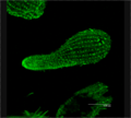
 MP-30 GFP
 -
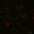
 Diatom chain
 -
 STD Depth Coded Stack Slices through Cells
 -
 Tetrachimena Beta Tubulin
 -
 Confocal measurement of 1-euro star 3d and euro
 -
 Confocal measurement of 1-euro star 3d Reflection
 -
 Depth Coded Phalloidin Stained Actin Filaments Cancer Cell
 -
 Mitotic spindle in Arabidopsis primary root meristem cells (anaphase)
 -
 Minski confocal patent figure 1
 -
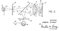
 Petran Patent Figure 2 cutout


Medical Disclaimer: WikiMD is for informational purposes only and is not a substitute for professional medical advice. Content may be inaccurate or outdated and should not be used for diagnosis or treatment. Always consult your healthcare provider for medical decisions. Verify information with trusted sources such as CDC.gov and NIH.gov. By using this site, you agree that WikiMD is not liable for any outcomes related to its content. See full disclaimer.
Credits:Most images are courtesy of Wikimedia commons, and templates, categories Wikipedia, licensed under CC BY SA or similar.
Translate page: - East Asian
中文,
日本,
한국어,
South Asian
हिन्दी,
தமிழ்,
తెలుగు,
Urdu,
ಕನ್ನಡ,
Southeast Asian
Indonesian,
Vietnamese,
Thai,
မြန်မာဘာသာ,
বাংলা
European
español,
Deutsch,
français,
Greek,
português do Brasil,
polski,
română,
русский,
Nederlands,
norsk,
svenska,
suomi,
Italian
Middle Eastern & African
عربى,
Turkish,
Persian,
Hebrew,
Afrikaans,
isiZulu,
Kiswahili,
Other
Bulgarian,
Hungarian,
Czech,
Swedish,
മലയാളം,
मराठी,
ਪੰਜਾਬੀ,
ગુજરાતી,
Portuguese,
Ukrainian
