Cotton wool spots
Cotton Wool Spots are small, white or grayish, opaque lesions on the retina that represent localized microinfarctions of the retinal nerve fiber layer. These spots are indicative of various systemic and ocular conditions, reflecting underlying abnormalities in retinal blood flow or retinal vascular integrity. Cotton wool spots are often associated with diseases that affect the vascular system, such as hypertension, diabetes mellitus, and HIV/AIDS, among others.
Etiology[edit]
Cotton wool spots can arise from a multitude of causes, primarily those that lead to retinal ischemia or disruption of axoplasmic flow within the retinal nerve fibers. The most common systemic conditions associated with these lesions include:
- Hypertension: Chronic high blood pressure can damage retinal blood vessels, leading to ischemia and cotton wool spot formation.
- Diabetes Mellitus: Diabetic retinopathy can cause microvascular damage and ischemia.
- HIV/AIDS: Infection with the human immunodeficiency virus can lead to immune suppression and various ocular manifestations, including cotton wool spots.
- Systemic Lupus Erythematosus (SLE): This autoimmune disease can affect multiple organ systems, including the eyes, leading to vascular changes and cotton wool spots.
- Giant Cell Arteritis: This is a form of vasculitis that can lead to occlusion of the retinal arteries and subsequent ischemia.
Pathophysiology[edit]
The pathogenesis of cotton wool spots involves the occlusion of precapillary arterioles, leading to infarction of the nerve fiber layer of the retina. This results in the accumulation of axoplasmic material, which appears ophthalmoscopically as fluffy white lesions. The size and shape of cotton wool spots can vary, but they typically measure less than a quarter of the optic disc diameter.
Clinical Presentation[edit]
Patients with cotton wool spots may be asymptomatic, with the lesions discovered incidentally during routine eye examination. In cases where the spots are extensive, individuals may experience visual disturbances, such as blurred vision or scotomas (blind spots). The presence of multiple cotton wool spots can indicate significant systemic disease, necessitating further medical evaluation and management.
Diagnosis[edit]
Diagnosis of cotton wool spots is primarily based on clinical examination using ophthalmoscopy or fundus photography. Fluorescein angiography may be employed to assess the retinal circulation and identify areas of non-perfusion. Optical coherence tomography (OCT) can also be useful in visualizing the retinal nerve fiber layer and confirming the diagnosis.
Management[edit]
The management of cotton wool spots involves addressing the underlying systemic condition. Control of systemic risk factors, such as blood pressure and blood sugar levels, is crucial. In cases related to infectious diseases or autoimmune conditions, appropriate systemic treatment is necessary. Regular follow-up with an ophthalmologist is important for monitoring the ocular manifestations and preventing potential complications.
Prognosis[edit]
The prognosis for patients with cotton wool spots largely depends on the underlying cause and the extent of systemic involvement. In many cases, the spots may resolve spontaneously once the systemic condition is adequately managed. However, persistent or recurrent lesions may indicate ongoing disease activity or inadequate control of systemic risk factors.
-
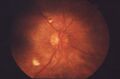 Cotton wool spots in proliferative retinopathy
 -
Cotton wool spots and hemorrhages


Medical Disclaimer: WikiMD is for informational purposes only and is not a substitute for professional medical advice. Content may be inaccurate or outdated and should not be used for diagnosis or treatment. Always consult your healthcare provider for medical decisions. Verify information with trusted sources such as CDC.gov and NIH.gov. By using this site, you agree that WikiMD is not liable for any outcomes related to its content. See full disclaimer.
Credits:Most images are courtesy of Wikimedia commons, and templates, categories Wikipedia, licensed under CC BY SA or similar.
Translate page: - East Asian
中文,
日本,
한국어,
South Asian
हिन्दी,
தமிழ்,
తెలుగు,
Urdu,
ಕನ್ನಡ,
Southeast Asian
Indonesian,
Vietnamese,
Thai,
မြန်မာဘာသာ,
বাংলা
European
español,
Deutsch,
français,
Greek,
português do Brasil,
polski,
română,
русский,
Nederlands,
norsk,
svenska,
suomi,
Italian
Middle Eastern & African
عربى,
Turkish,
Persian,
Hebrew,
Afrikaans,
isiZulu,
Kiswahili,
Other
Bulgarian,
Hungarian,
Czech,
Swedish,
മലയാളം,
मराठी,
ਪੰਜਾਬੀ,
ગુજરાતી,
Portuguese,
Ukrainian
