Craniofacial cleft
Craniofacial cleft is a congenital deformity characterized by an abnormal gap or cleft in the face and/or the cranium. These clefts can vary greatly in size, shape, and location, affecting not only the appearance and function of the face but also potentially impacting the brain and the development of the skull. Craniofacial clefts are part of a broader category of craniofacial anomalies and can occur as isolated defects or as part of a syndrome.
Etiology[edit]
The exact cause of craniofacial clefts is not fully understood, but it is believed to be a combination of genetic and environmental factors. Disruptions in the normal development of the face and skull during embryonic growth lead to these anomalies. Factors that may increase the risk include genetic mutations, maternal smoking, use of certain medications during pregnancy, and nutritional deficiencies.
Classification[edit]
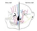
Craniofacial clefts are classified according to their location and the structures they involve. The most widely used classification system is the Tessier cleft classification system, which numbers clefts from 0 to 14, based on their anatomical position. This system helps in understanding the complexity of the clefts and planning for surgical intervention.
Symptoms and Complications[edit]
The symptoms of craniofacial clefts can vary widely depending on the severity and location of the cleft. Common issues include:
- Difficulty with feeding and swallowing
- Breathing problems
- Speech difficulties
- Hearing loss
- Dental problems
- Visual impairments
Complications may also arise from associated anomalies, such as heart defects, brain abnormalities, and limb deformities in syndromic cases.
Diagnosis[edit]
Diagnosis of craniofacial clefts typically occurs through prenatal imaging, such as ultrasound, and is confirmed after birth by physical examination. Advanced imaging techniques, like CT scans and MRI, are used to assess the extent of the cleft and plan for surgical correction.
Treatment[edit]
Treatment of craniofacial clefts is complex and often requires a multidisciplinary approach, involving surgeons, pediatricians, dentists, speech therapists, and other specialists. The primary goal of treatment is to improve function and appearance. This often involves multiple surgeries over the course of several years, including reconstructive surgery to close the cleft, orthodontic treatment, and speech therapy.
Prognosis[edit]
The prognosis for individuals with craniofacial clefts varies depending on the severity of the cleft and the presence of associated anomalies. With early and comprehensive treatment, many individuals can lead healthy, fulfilling lives.
See Also[edit]
This medical article is a stub. You can help WikiMD by expanding the page. |
-
 Craniofacial cleft
 -
 Craniofacial cleft
 -
 Craniofacial cleft
 -
 Craniofacial cleft
 -
 Craniofacial cleft
 -
 Craniofacial cleft
 -
 Craniofacial cleft
 -
 Craniofacial cleft
 -
 Craniofacial cleft


Medical Disclaimer: WikiMD is for informational purposes only and is not a substitute for professional medical advice. Content may be inaccurate or outdated and should not be used for diagnosis or treatment. Always consult your healthcare provider for medical decisions. Verify information with trusted sources such as CDC.gov and NIH.gov. By using this site, you agree that WikiMD is not liable for any outcomes related to its content. See full disclaimer.
Credits:Most images are courtesy of Wikimedia commons, and templates, categories Wikipedia, licensed under CC BY SA or similar.
Translate page: - East Asian
中文,
日本,
한국어,
South Asian
हिन्दी,
தமிழ்,
తెలుగు,
Urdu,
ಕನ್ನಡ,
Southeast Asian
Indonesian,
Vietnamese,
Thai,
မြန်မာဘာသာ,
বাংলা
European
español,
Deutsch,
français,
Greek,
português do Brasil,
polski,
română,
русский,
Nederlands,
norsk,
svenska,
suomi,
Italian
Middle Eastern & African
عربى,
Turkish,
Persian,
Hebrew,
Afrikaans,
isiZulu,
Kiswahili,
Other
Bulgarian,
Hungarian,
Czech,
Swedish,
മലയാളം,
मराठी,
ਪੰਜਾਬੀ,
ગુજરાતી,
Portuguese,
Ukrainian
