Ductus reuniens
Anatomical structure in the inner ear
The ductus reuniens is a small but significant anatomical structure within the inner ear. It plays a crucial role in the auditory and vestibular systems by connecting the cochlea and the saccule, two essential components of the inner ear. Understanding the ductus reuniens is important for comprehending how the inner ear functions in hearing and balance.
Anatomy[edit]
The ductus reuniens is a narrow duct that forms part of the membranous labyrinth of the inner ear. It is located within the temporal bone, which houses the structures of the ear. The ductus reuniens connects the cochlear duct of the cochlea to the saccule, which is one of the two otolithic organs in the vestibular system.
Structure[edit]
The ductus reuniens is a slender tube that measures only a few millimeters in length. It is lined with a thin layer of epithelial cells and is filled with endolymph, a fluid that is crucial for the proper functioning of the inner ear. The ductus reuniens is part of the continuous endolymphatic system that includes the cochlear duct, the saccule, and the utricle.
Location[edit]
The ductus reuniens is situated in the petrous part of the temporal bone, which is the hardest bone in the human body. It lies in close proximity to other important structures of the inner ear, including the semicircular canals and the vestibular nerve.
Function[edit]
The primary function of the ductus reuniens is to facilitate the flow of endolymph between the cochlea and the saccule. This connection is vital for maintaining the ionic composition and pressure of the endolymph, which is necessary for the transduction of sound waves into nerve impulses in the cochlea and for the detection of linear acceleration in the saccule.
Role in Hearing[edit]
In the process of hearing, sound waves enter the ear and are transmitted to the cochlea, where they cause the endolymph to move. This movement stimulates the hair cells in the cochlea, leading to the generation of nerve impulses that are sent to the brain via the auditory nerve. The ductus reuniens ensures that the endolymphatic fluid is properly balanced, which is essential for accurate sound perception.
Role in Balance[edit]
The saccule, connected to the cochlea via the ductus reuniens, is involved in the vestibular system, which helps maintain balance and spatial orientation. The movement of endolymph within the saccule, influenced by gravity and linear acceleration, stimulates hair cells that send signals to the brain about the body's position and movement.
Clinical Significance[edit]
Disorders affecting the ductus reuniens can lead to hearing and balance problems. Conditions such as Ménière's disease may involve dysfunction of the endolymphatic system, including the ductus reuniens, resulting in symptoms like vertigo, tinnitus, and hearing loss.
Related pages[edit]
-
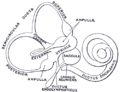 Gray's Anatomy illustration of the ductus reuniens


Medical Disclaimer: WikiMD is for informational purposes only and is not a substitute for professional medical advice. Content may be inaccurate or outdated and should not be used for diagnosis or treatment. Always consult your healthcare provider for medical decisions. Verify information with trusted sources such as CDC.gov and NIH.gov. By using this site, you agree that WikiMD is not liable for any outcomes related to its content. See full disclaimer.
Credits:Most images are courtesy of Wikimedia commons, and templates, categories Wikipedia, licensed under CC BY SA or similar.
Translate page: - East Asian
中文,
日本,
한국어,
South Asian
हिन्दी,
தமிழ்,
తెలుగు,
Urdu,
ಕನ್ನಡ,
Southeast Asian
Indonesian,
Vietnamese,
Thai,
မြန်မာဘာသာ,
বাংলা
European
español,
Deutsch,
français,
Greek,
português do Brasil,
polski,
română,
русский,
Nederlands,
norsk,
svenska,
suomi,
Italian
Middle Eastern & African
عربى,
Turkish,
Persian,
Hebrew,
Afrikaans,
isiZulu,
Kiswahili,
Other
Bulgarian,
Hungarian,
Czech,
Swedish,
മലയാളം,
मराठी,
ਪੰਜਾਬੀ,
ગુજરાતી,
Portuguese,
Ukrainian
