Elbow
The elbow is a complex hinge joint formed by the articulation of three bones: the humerus, radius, and ulna. It is a fundamental part of the human skeletal system and plays a crucial role in upper limb movement.
Anatomy[edit]
The elbow joint is where the long bone at the top of your arm, known as the humerus, meets the two bones in your forearm, known as the radius and ulna. It is one of the largest joints in the body and is surrounded by muscles, ligaments, and tendons that enable a wide range of movements.
Bones[edit]
- Humerus: The humerus is the long bone in the upper arm. It has two articulating surfaces that interact with the radius and ulna to form the elbow joint.
- Radius: The radius is one of the two bones in the forearm. It is located on the thumb side of the arm and rotates to allow the hand to turn palm up.
- Ulna: The ulna is the other bone in the forearm, located on the pinky side. It has a hook-like process, the olecranon, which forms the bony prominence of the elbow.
Ligaments and Tendons[edit]
The elbow joint is supported by several ligaments and tendons, including the annular ligament, the radial collateral ligament, the ulnar collateral ligament, and the quadrate ligament.
Function[edit]
The elbow joint allows for two basic movements: flexion and extension. Flexion occurs when the angle of the joint decreases, as in bending the elbow to bring the hand closer to the shoulder. Extension is the opposite movement, increasing the angle of the joint to straighten the arm.
Conditions and Disorders[edit]
There are many conditions and disorders that can affect the elbow, including tennis elbow, golfer's elbow, bursitis, arthritis, and fractures. These conditions can cause pain, swelling, stiffness, and limit the range of motion of the elbow.
Treatment[edit]
Treatment for elbow conditions and disorders depends on the specific diagnosis and may include rest, physical therapy, medication, and in some cases, surgery.
See Also[edit]

This WikiMD article can only be edited by registered and verified editors. You can log in or register.
-
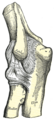
 Elbow
Elbow -
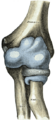
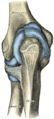
 Diagram of the elbow joint
 -
 Bones of the right arm. Anterior aspect.
 -
 Bones of the right arm. Posterior aspect.
 -
 The right humerus. Anterior view.
 -
 The right humerus. Posterior view.
 -
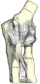
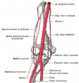 The ligaments of the elbow joint. Anterior view.
 -
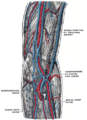 The supinator.
 -
 Elbow
Elbow -
 Elbow
Elbow -
 Elbow
Elbow -
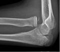 X-ray of ventral dislocation of the radial head with calcification of annular ligament


Medical Disclaimer: WikiMD is for informational purposes only and is not a substitute for professional medical advice. Content may be inaccurate or outdated and should not be used for diagnosis or treatment. Always consult your healthcare provider for medical decisions. Verify information with trusted sources such as CDC.gov and NIH.gov. By using this site, you agree that WikiMD is not liable for any outcomes related to its content. See full disclaimer.
Credits:Most images are courtesy of Wikimedia commons, and templates, categories Wikipedia, licensed under CC BY SA or similar.
Translate page: - East Asian
中文,
日本,
한국어,
South Asian
हिन्दी,
தமிழ்,
తెలుగు,
Urdu,
ಕನ್ನಡ,
Southeast Asian
Indonesian,
Vietnamese,
Thai,
မြန်မာဘာသာ,
বাংলা
European
español,
Deutsch,
français,
Greek,
português do Brasil,
polski,
română,
русский,
Nederlands,
norsk,
svenska,
suomi,
Italian
Middle Eastern & African
عربى,
Turkish,
Persian,
Hebrew,
Afrikaans,
isiZulu,
Kiswahili,
Other
Bulgarian,
Hungarian,
Czech,
Swedish,
മലയാളം,
मराठी,
ਪੰਜਾਬੀ,
ગુજરાતી,
Portuguese,
Ukrainian
