Female reproductive system
Female reproductive system is a complex system in the female body that plays a crucial role in reproduction. It includes various organs and structures such as the ovaries, fallopian tubes, uterus, vagina, and vulva.
Anatomy[edit]
The female reproductive system is made up of internal and external structures. The internal structures include the ovaries, fallopian tubes, uterus, and vagina. The external structures include the vulva, which consists of the labia majora, labia minora, clitoris, and vaginal opening.
Ovaries[edit]
The ovaries are a pair of small, almond-shaped organs located on either side of the uterus. They are responsible for producing eggs (or oocytes) and the female hormones estrogen and progesterone.
Fallopian Tubes[edit]
The fallopian tubes are a pair of tubes that connect the ovaries to the uterus. They are the site where fertilization usually occurs.
Uterus[edit]
The uterus, also known as the womb, is a pear-shaped organ located in the lower abdomen. It is where the fetus develops during pregnancy.
Vagina[edit]
The vagina is a muscular canal that extends from the cervix, which is the lower part of the uterus, to the external part of the female genitalia, which is called the vulva.
Function[edit]
The main function of the female reproductive system is to produce eggs for fertilization and to provide a place where a fertilized egg can develop into a fetus. The ovaries produce the female eggs or oocytes. If an oocyte is fertilized by a sperm, it will move into the uterus and implant itself into the uterine wall, where it will develop into a fetus.
Diseases and Conditions[edit]
There are many diseases and conditions that can affect the female reproductive system. These include, but are not limited to, endometriosis, polycystic ovary syndrome, uterine fibroids, and various forms of gynecological cancer.
See Also[edit]

This WikiMD article can only be edited by registered and verified editors. You can log in or register.
-
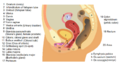 Lateral view of the female reproductive system
 -
Diagram of the female reproductive system with numbered labels
-
Female reproductive system
-
Diagram showing the vaginal opening
-
Illustration of the female reproductive system
-
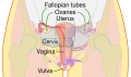 Diagram of the female reproductive system


Medical Disclaimer: WikiMD is for informational purposes only and is not a substitute for professional medical advice. Content may be inaccurate or outdated and should not be used for diagnosis or treatment. Always consult your healthcare provider for medical decisions. Verify information with trusted sources such as CDC.gov and NIH.gov. By using this site, you agree that WikiMD is not liable for any outcomes related to its content. See full disclaimer.
Credits:Most images are courtesy of Wikimedia commons, and templates, categories Wikipedia, licensed under CC BY SA or similar.
Translate page: - East Asian
中文,
日本,
한국어,
South Asian
हिन्दी,
தமிழ்,
తెలుగు,
Urdu,
ಕನ್ನಡ,
Southeast Asian
Indonesian,
Vietnamese,
Thai,
မြန်မာဘာသာ,
বাংলা
European
español,
Deutsch,
français,
Greek,
português do Brasil,
polski,
română,
русский,
Nederlands,
norsk,
svenska,
suomi,
Italian
Middle Eastern & African
عربى,
Turkish,
Persian,
Hebrew,
Afrikaans,
isiZulu,
Kiswahili,
Other
Bulgarian,
Hungarian,
Czech,
Swedish,
മലയാളം,
मराठी,
ਪੰਜਾਬੀ,
ગુજરાતી,
Portuguese,
Ukrainian
