Sella turcica
(Redirected from Fossa hypophyseos)
Saddle-shaped depression in the sphenoid bone of the human skull
| Sella turcica | |
|---|---|
| Sella turcica.jpg | |
| Sella turcica shown in red | |
| General Information | |
| Latin | |
| Greek | |
| TA98 | |
| TA2 | |
| FMA | |
| Details | |
| System | |
| Artery | |
| Vein | |
| Nerve | |
| Lymphatic drainage | |
| Precursor | |
| Function | |
| Identifiers | |
| Clinical significance | |
| Notes | |
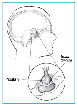
The sella turcica (Latin for "Turkish saddle") is a saddle-shaped depression in the body of the sphenoid bone of the human skull. It is located in the middle cranial fossa, a central depression in the floor of the cranial cavity. The sella turcica is an important anatomical landmark because it houses the pituitary gland.
Anatomy[edit | edit source]
The sella turcica is bounded by the tuberculum sellae anteriorly and the dorsum sellae posteriorly. The hypophyseal fossa, the deepest part of the sella turcica, contains the pituitary gland. The sella turcica is covered by a dural fold called the diaphragma sellae, which has an opening for the passage of the pituitary stalk.
Function[edit | edit source]
The primary function of the sella turcica is to protect and support the pituitary gland. The pituitary gland, also known as the hypophysis, is a crucial endocrine gland that regulates various physiological processes through the secretion of hormones.
Clinical significance[edit | edit source]
Abnormalities in the size or shape of the sella turcica can be indicative of various medical conditions. For example, an enlarged sella turcica may be associated with a pituitary adenoma, a benign tumor of the pituitary gland. Other conditions that can affect the sella turcica include empty sella syndrome, where the sella turcica appears empty on imaging due to the flattening of the pituitary gland, and craniopharyngioma, a type of brain tumor that can affect the region.
Imaging[edit | edit source]
The sella turcica can be visualized using various imaging techniques, including X-ray, computed tomography (CT), and magnetic resonance imaging (MRI). These imaging modalities are essential for diagnosing abnormalities of the pituitary gland and surrounding structures.
See also[edit | edit source]
References[edit | edit source]
Search WikiMD
Ad.Tired of being Overweight? Try W8MD's NYC physician weight loss.
Semaglutide (Ozempic / Wegovy and Tirzepatide (Mounjaro / Zepbound) available. Call 718 946 5500.
Advertise on WikiMD
|
WikiMD's Wellness Encyclopedia |
| Let Food Be Thy Medicine Medicine Thy Food - Hippocrates |
Translate this page: - East Asian
中文,
日本,
한국어,
South Asian
हिन्दी,
தமிழ்,
తెలుగు,
Urdu,
ಕನ್ನಡ,
Southeast Asian
Indonesian,
Vietnamese,
Thai,
မြန်မာဘာသာ,
বাংলা
European
español,
Deutsch,
français,
Greek,
português do Brasil,
polski,
română,
русский,
Nederlands,
norsk,
svenska,
suomi,
Italian
Middle Eastern & African
عربى,
Turkish,
Persian,
Hebrew,
Afrikaans,
isiZulu,
Kiswahili,
Other
Bulgarian,
Hungarian,
Czech,
Swedish,
മലയാളം,
मराठी,
ਪੰਜਾਬੀ,
ગુજરાતી,
Portuguese,
Ukrainian
Medical Disclaimer: WikiMD is not a substitute for professional medical advice. The information on WikiMD is provided as an information resource only, may be incorrect, outdated or misleading, and is not to be used or relied on for any diagnostic or treatment purposes. Please consult your health care provider before making any healthcare decisions or for guidance about a specific medical condition. WikiMD expressly disclaims responsibility, and shall have no liability, for any damages, loss, injury, or liability whatsoever suffered as a result of your reliance on the information contained in this site. By visiting this site you agree to the foregoing terms and conditions, which may from time to time be changed or supplemented by WikiMD. If you do not agree to the foregoing terms and conditions, you should not enter or use this site. See full disclaimer.
Credits:Most images are courtesy of Wikimedia commons, and templates, categories Wikipedia, licensed under CC BY SA or similar.
Contributors: Prab R. Tumpati, MD


