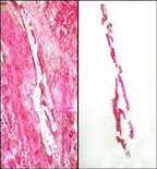
Laser capture microdissection
Laser Capture Microdissection (LCM) is a technique used in the field of microscopy to selectively isolate specific cells from a heterogeneous tissue section or cell population under direct microscopic visualization. LCM technology enables researchers to precisely select and isolate cells of interest from a wide variety of specimens, including tissue sections, smear preparations, and cell cultures, by using a focused laser beam. This method is crucial for the study of cell biology, genomics, proteomics, and molecular pathology, as it allows for the extraction of high-quality DNA, RNA, and proteins from specific cell populations, thereby facilitating detailed molecular analyses.
History[edit | edit source]
The concept of LCM was first introduced in the late 1990s as a method to address the need for a precise and efficient way to isolate specific cells from complex tissues. The development of LCM technology represented a significant advancement in the field of molecular biology, as it provided a solution to the challenge of studying individual cell types within mixed cell populations.
Principle[edit | edit source]
LCM operates on the principle of using a laser beam to selectively adhere targeted cells to a transfer film, which is then lifted away from the tissue section. This process allows for the isolation of pure cell populations without contamination from surrounding cells. The procedure can be performed under direct visualization, enabling the operator to accurately select the desired cells.
Applications[edit | edit source]
LCM has a wide range of applications in biomedical research, including:
- Cancer research: Isolating tumor cells from surrounding stromal and immune cells to study cancer genetics and tumor microenvironments.
- Neuroscience: Separating specific neuron types for the study of neurological diseases and brain functions.
- Developmental biology: Analyzing spatial and temporal gene expression patterns in developing tissues.
- Pathology: Enabling molecular analysis of diseased tissues, including infectious diseases and genetic disorders.
Procedure[edit | edit source]
The LCM procedure involves several steps: 1. Preparation of tissue sections or cell samples on microscope slides. 2. Staining of samples to identify cells of interest, while preserving the integrity of nucleic acids and proteins. 3. Microscopic visualization of the sample to locate and target specific cells. 4. Activation of the laser to capture targeted cells onto a transfer film. 5. Extraction of captured cells for subsequent molecular analysis.
Advantages[edit | edit source]
LCM offers several advantages over traditional cell isolation techniques:
- Precision: Allows for the isolation of specific cell types from complex tissues.
- Purity: Ensures high purity of isolated cells, minimizing contamination.
- Versatility: Applicable to a wide range of tissue types and sample preparations.
- Preservation: Maintains the integrity of nucleic acids and proteins for downstream analyses.
Challenges[edit | edit source]
Despite its advantages, LCM faces some challenges:
- Technical complexity: Requires specialized equipment and skilled operation.
- Sample preparation: Necessitates careful preparation to preserve molecular integrity.
- Sensitivity: Limited by the sensitivity of downstream molecular analyses.
Future Directions[edit | edit source]
The future of LCM lies in the integration with advanced molecular analysis techniques, such as next-generation sequencing and mass spectrometry, to enhance our understanding of cellular functions and disease mechanisms at an unprecedented resolution.
| This article is a stub. You can help WikiMD by registering to expand it. |
Laser_capture_microdissection[edit | edit source]
Search WikiMD
Ad.Tired of being Overweight? Try W8MD's physician weight loss program.
Semaglutide (Ozempic / Wegovy and Tirzepatide (Mounjaro / Zepbound) available.
Advertise on WikiMD
|
WikiMD's Wellness Encyclopedia |
| Let Food Be Thy Medicine Medicine Thy Food - Hippocrates |
Translate this page: - East Asian
中文,
日本,
한국어,
South Asian
हिन्दी,
தமிழ்,
తెలుగు,
Urdu,
ಕನ್ನಡ,
Southeast Asian
Indonesian,
Vietnamese,
Thai,
မြန်မာဘာသာ,
বাংলা
European
español,
Deutsch,
français,
Greek,
português do Brasil,
polski,
română,
русский,
Nederlands,
norsk,
svenska,
suomi,
Italian
Middle Eastern & African
عربى,
Turkish,
Persian,
Hebrew,
Afrikaans,
isiZulu,
Kiswahili,
Other
Bulgarian,
Hungarian,
Czech,
Swedish,
മലയാളം,
मराठी,
ਪੰਜਾਬੀ,
ગુજરાતી,
Portuguese,
Ukrainian
Medical Disclaimer: WikiMD is not a substitute for professional medical advice. The information on WikiMD is provided as an information resource only, may be incorrect, outdated or misleading, and is not to be used or relied on for any diagnostic or treatment purposes. Please consult your health care provider before making any healthcare decisions or for guidance about a specific medical condition. WikiMD expressly disclaims responsibility, and shall have no liability, for any damages, loss, injury, or liability whatsoever suffered as a result of your reliance on the information contained in this site. By visiting this site you agree to the foregoing terms and conditions, which may from time to time be changed or supplemented by WikiMD. If you do not agree to the foregoing terms and conditions, you should not enter or use this site. See full disclaimer.
Credits:Most images are courtesy of Wikimedia commons, and templates, categories Wikipedia, licensed under CC BY SA or similar.
Contributors: Prab R. Tumpati, MD


