Microtome
Microtome is a tool used in microscopy to cut extremely thin slices of material, known as sections. They are an important device in medical research, forensic investigation, and in the diagnosis of diseases. The slices are often stained and then examined under a microscope.
History[edit]
The earliest devices were simply a sharp knife and a hand, but the development of the microtome led to the cutting of tissues into thin sections for microscopic examination. The first microtomes were made in 1770 by George Adams, Jr. and further developed in 1835 by Andrew Prichard.
Types of Microtomes[edit]
There are several types of microtomes, including the rotary microtome, cryomicrotome, ultramicrotome, vibrating microtome, and laser microtome. Each type has its own advantages and disadvantages, and is used for different purposes.
Uses[edit]
Microtomes are used in both clinical and research settings to prepare slides of tissue for viewing under a microscope. These slides can be used to diagnose diseases, such as cancer, and to research the effects of diseases on tissue structure.
See Also[edit]

This WikiMD article can only be edited by registered and verified editors. You can log in or register.
Microtome[edit]
-
Cummings 1774 Microtome
-
Anonymous 1910 An Eighteenth Century Microtome
-
Microtome 1905
-
Sledge Microtome
-
Microtome 1
-
Microtome Principle
-
 Cryostat Microtome
 -
Microtome Ultras
-
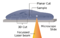 Laser Microtome Schematic
 -
Diamond Knife Blade Edge
-
Одноразовое лезвие для микротома
-
 Microtome Knife Profile


Medical Disclaimer: WikiMD is for informational purposes only and is not a substitute for professional medical advice. Content may be inaccurate or outdated and should not be used for diagnosis or treatment. Always consult your healthcare provider for medical decisions. Verify information with trusted sources such as CDC.gov and NIH.gov. By using this site, you agree that WikiMD is not liable for any outcomes related to its content. See full disclaimer.
Credits:Most images are courtesy of Wikimedia commons, and templates, categories Wikipedia, licensed under CC BY SA or similar.
Translate page: - East Asian
中文,
日本,
한국어,
South Asian
हिन्दी,
தமிழ்,
తెలుగు,
Urdu,
ಕನ್ನಡ,
Southeast Asian
Indonesian,
Vietnamese,
Thai,
မြန်မာဘာသာ,
বাংলা
European
español,
Deutsch,
français,
Greek,
português do Brasil,
polski,
română,
русский,
Nederlands,
norsk,
svenska,
suomi,
Italian
Middle Eastern & African
عربى,
Turkish,
Persian,
Hebrew,
Afrikaans,
isiZulu,
Kiswahili,
Other
Bulgarian,
Hungarian,
Czech,
Swedish,
മലയാളം,
मराठी,
ਪੰਜਾਬੀ,
ગુજરાતી,
Portuguese,
Ukrainian
