Owl's eye appearance
Owl's eye appearance refers to a distinctive radiological and histopathological finding characterized by large, inclusion-bearing cells that resemble the eyes of an owl. This appearance is most commonly associated with infections caused by the Cytomegalovirus (CMV) but can also be seen in other conditions. The term is used descriptively in medical diagnostics and is significant in the fields of Pathology and Radiology.
Etiology[edit]
The owl's eye appearance is primarily linked to Cytomegalovirus (CMV) infections. CMV is a type of herpesvirus that can cause disease in fetuses, neonates, and immunocompromised individuals such as transplant recipients and patients with HIV/AIDS. The virus infects cells and enlarges them, leading to the characteristic appearance. Besides CMV, other conditions can occasionally produce similar imaging or histological patterns, though they are less common.
Pathophysiology[edit]
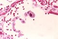
In the context of CMV infection, the owl's eye appearance arises from the viral cytopathic effect. The virus induces considerable enlargement of the infected cells, and within these cells, intranuclear inclusions are formed. These inclusions push the cell's chromatin to the periphery, creating a clear zone that resembles an owl's eye. This effect is most evident in histopathological examinations of affected tissues.
Clinical Significance[edit]
The identification of an owl's eye appearance can be crucial for diagnosing CMV infections, especially in patients with unclear clinical presentations. In immunocompromised patients, early detection and treatment of CMV can significantly affect the prognosis. Therefore, recognizing this pattern on histopathology or imaging studies can guide clinicians towards more specific diagnostic tests for CMV and appropriate antiviral therapy.
Diagnosis[edit]
Diagnosis of conditions associated with the owl's eye appearance involves a combination of clinical evaluation, imaging studies, and laboratory tests. Histopathological examination of biopsy samples is the gold standard for observing the owl's eye appearance directly. In addition to tissue biopsy, polymerase chain reaction (PCR) testing for CMV DNA in blood or other body fluids can support the diagnosis.
Treatment[edit]
Treatment for CMV infections, the most common cause of the owl's eye appearance, includes antiviral medications such as ganciclovir, valganciclovir, foscarnet, and cidofovir. The choice of treatment depends on the severity of the infection, the patient's immune status, and the presence of drug-resistant CMV strains. Early and appropriate antiviral therapy is essential for managing CMV infections effectively.
Conclusion[edit]
The owl's eye appearance is a distinctive finding associated with CMV infections and other conditions. Recognizing this pattern can be crucial for the timely diagnosis and treatment of affected patients, particularly those who are immunocompromised. As such, it remains an important aspect of diagnostic pathology and radiology.
This medical article is a stub. You can help WikiMD by expanding the page. |
Owl's_eye_appearance[edit]
-
Orange eye
-
 Cytomegalovirus inclusion
 -
CMV neuronal inclusions


Medical Disclaimer: WikiMD is for informational purposes only and is not a substitute for professional medical advice. Content may be inaccurate or outdated and should not be used for diagnosis or treatment. Always consult your healthcare provider for medical decisions. Verify information with trusted sources such as CDC.gov and NIH.gov. By using this site, you agree that WikiMD is not liable for any outcomes related to its content. See full disclaimer.
Credits:Most images are courtesy of Wikimedia commons, and templates, categories Wikipedia, licensed under CC BY SA or similar.
Translate page: - East Asian
中文,
日本,
한국어,
South Asian
हिन्दी,
தமிழ்,
తెలుగు,
Urdu,
ಕನ್ನಡ,
Southeast Asian
Indonesian,
Vietnamese,
Thai,
မြန်မာဘာသာ,
বাংলা
European
español,
Deutsch,
français,
Greek,
português do Brasil,
polski,
română,
русский,
Nederlands,
norsk,
svenska,
suomi,
Italian
Middle Eastern & African
عربى,
Turkish,
Persian,
Hebrew,
Afrikaans,
isiZulu,
Kiswahili,
Other
Bulgarian,
Hungarian,
Czech,
Swedish,
മലയാളം,
मराठी,
ਪੰਜਾਬੀ,
ગુજરાતી,
Portuguese,
Ukrainian

.jpg)