PAS diastase stain
PAS diastase stain, also known as Periodic acid-Schiff with diastase stain, is a histochemical staining method used in pathology and histology to identify complex carbohydrates in tissues. This technique is particularly useful for highlighting structures such as glycogen, mucosubstances, and fungal cell walls. The PAS diastase stain combines the Periodic acid-Schiff (PAS) reaction with diastase treatment to specifically target glycogen, making it a critical tool in both diagnostic pathology and research.
Overview[edit]
The PAS diastase stain procedure involves the application of periodic acid, which oxidizes the carbon-carbon bonds in carbohydrates, creating aldehyde groups. These aldehyde groups then react with Schiff reagent, resulting in a magenta color. The addition of diastase, an enzyme that digests glycogen, before the PAS reaction allows for the differentiation between glycogen and other PAS-positive substances. Structures that lose their magenta color after diastase treatment are identified as glycogen, whereas those that retain the color are typically other types of polysaccharides or mucosubstances.
Applications[edit]
The PAS diastase stain is widely used in the diagnosis of various diseases and conditions. In Liver pathology, it helps in identifying glycogen storage diseases by demonstrating abnormal glycogen accumulation within hepatocytes. In Dermatopathology, it is used to detect fungal infections, as the cell walls of fungi are rich in polysaccharides that are PAS-positive. The stain is also employed in the diagnosis of Alimentary tract pathology, particularly in identifying conditions like Whipple's disease, where PAS-positive macrophages are a hallmark feature.
Procedure[edit]
The staining procedure involves several steps:
- Fixation of the tissue sample, typically in formalin.
- Embedding the tissue in paraffin and sectioning.
- Deparaffinization and hydration of tissue sections.
- Treatment with diastase enzyme to digest glycogen.
- Oxidation with periodic acid.
- Reaction with Schiff reagent.
- Counterstaining, often with hematoxylin, to provide a background contrast.
- Dehydration, clearing, and mounting of the tissue sections for microscopic examination.
Interpretation[edit]
Under the microscope, PAS-positive substances appear magenta or purple, while the background and other tissue components are counterstained blue by hematoxylin. The presence or absence of magenta staining after diastase treatment helps in identifying glycogen versus other substances.
Limitations[edit]
While the PAS diastase stain is a powerful tool, it has limitations. The specificity of the stain can be affected by the fixation and processing of the tissue. Overdigestion or underdigestion with diastase can lead to false-negative or false-positive results, respectively. Therefore, careful control of staining conditions and interpretation by experienced pathologists is essential.
See Also[edit]
This medical article is a stub. You can help WikiMD by expanding the page. |
-
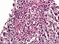 Histoplasma in granuloma stained with PAS diastase
 -
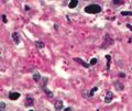 Histoplasma stained with PAS diastase


Medical Disclaimer: WikiMD is for informational purposes only and is not a substitute for professional medical advice. Content may be inaccurate or outdated and should not be used for diagnosis or treatment. Always consult your healthcare provider for medical decisions. Verify information with trusted sources such as CDC.gov and NIH.gov. By using this site, you agree that WikiMD is not liable for any outcomes related to its content. See full disclaimer.
Credits:Most images are courtesy of Wikimedia commons, and templates, categories Wikipedia, licensed under CC BY SA or similar.
Translate page: - East Asian
中文,
日本,
한국어,
South Asian
हिन्दी,
தமிழ்,
తెలుగు,
Urdu,
ಕನ್ನಡ,
Southeast Asian
Indonesian,
Vietnamese,
Thai,
မြန်မာဘာသာ,
বাংলা
European
español,
Deutsch,
français,
Greek,
português do Brasil,
polski,
română,
русский,
Nederlands,
norsk,
svenska,
suomi,
Italian
Middle Eastern & African
عربى,
Turkish,
Persian,
Hebrew,
Afrikaans,
isiZulu,
Kiswahili,
Other
Bulgarian,
Hungarian,
Czech,
Swedish,
മലയാളം,
मराठी,
ਪੰਜਾਬੀ,
ગુજરાતી,
Portuguese,
Ukrainian
