Resection margin
Resection margin refers to the edge or boundary of a surgically removed tissue, typically analyzed in the context of cancer surgery. The primary goal of examining the resection margin is to determine whether the surgery successfully removed all cancerous cells, which is crucial for assessing the likelihood of cancer recurrence and the need for additional treatments. Resection margins are categorized as either clear (negative), close, or involved (positive), based on the presence or absence of cancer cells at the edge of the removed tissue.
Definition and Importance[edit]
The resection margin is the boundary of tissue removed during surgery, which is examined microscopically to assess if it contains any cancer cells. A clear margin indicates no cancer cells are found at the edge, suggesting that the tumor has been fully excised. An involved margin means that cancer cells are present at the edge, indicating that some of the tumor may have been left behind. A close margin is when cancer cells are found near the edge, but not at the very boundary, posing a dilemma regarding the adequacy of tumor removal.
Types of Resection Margins[edit]
- Radial Margin: Particularly relevant in the resection of tubular structures like the esophagus or rectum, where the margin is assessed around the circumference of the tube.
- Deep Margin: The margin farthest from the body surface, important in surgeries where the depth of tumor invasion is a concern.
- Superficial Margin: The surface closest to the skin or body cavity, significant in surgeries for tumors near the body surface.
Clinical Significance[edit]
The status of the resection margin is a critical factor in determining the need for additional treatments such as chemotherapy, radiation therapy, or further surgical intervention. Positive margins often necessitate additional therapy to manage the risk of recurrence, while clear margins may indicate that the surgical removal was likely curative.
Pathological Examination[edit]
Pathologists play a crucial role in evaluating resection margins. The process involves careful examination of the surgically removed tissue, staining, and microscopic analysis to identify the presence of cancer cells at the margins. This assessment helps guide post-surgical treatment decisions and prognosis.
Challenges and Considerations[edit]
Achieving clear margins can be challenging, especially in cases where the tumor is located near critical structures or has irregular boundaries. Surgeons must balance the goal of complete tumor removal with the preservation of normal tissue function. In some cases, preoperative treatments such as chemotherapy or radiation therapy may be used to shrink the tumor and facilitate clear margins.
Future Directions[edit]
Advancements in surgical techniques, imaging technologies, and molecular diagnostics are contributing to improved accuracy in achieving and assessing clear resection margins. Intraoperative imaging and frozen section analysis are examples of techniques that can help surgeons achieve better outcomes.
This medical article is a stub. You can help WikiMD by expanding the page. |
-
Schematic showing positive margin and negative margin with English notations
-
Myopericytoma - intermediate magnification
-
Positive margin with cautery artefact - adenocarcinoma - low magnification
-
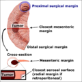 Edges and margins in intestinal tumor


Medical Disclaimer: WikiMD is for informational purposes only and is not a substitute for professional medical advice. Content may be inaccurate or outdated and should not be used for diagnosis or treatment. Always consult your healthcare provider for medical decisions. Verify information with trusted sources such as CDC.gov and NIH.gov. By using this site, you agree that WikiMD is not liable for any outcomes related to its content. See full disclaimer.
Credits:Most images are courtesy of Wikimedia commons, and templates, categories Wikipedia, licensed under CC BY SA or similar.
Translate page: - East Asian
中文,
日本,
한국어,
South Asian
हिन्दी,
தமிழ்,
తెలుగు,
Urdu,
ಕನ್ನಡ,
Southeast Asian
Indonesian,
Vietnamese,
Thai,
မြန်မာဘာသာ,
বাংলা
European
español,
Deutsch,
français,
Greek,
português do Brasil,
polski,
română,
русский,
Nederlands,
norsk,
svenska,
suomi,
Italian
Middle Eastern & African
عربى,
Turkish,
Persian,
Hebrew,
Afrikaans,
isiZulu,
Kiswahili,
Other
Bulgarian,
Hungarian,
Czech,
Swedish,
മലയാളം,
मराठी,
ਪੰਜਾਬੀ,
ગુજરાતી,
Portuguese,
Ukrainian
