Tympanic duct
Part of the inner ear involved in hearing
The tympanic duct, also known as the scala tympani, is one of the three fluid-filled passages in the cochlea of the inner ear. It plays a crucial role in the process of hearing by transmitting sound vibrations from the oval window to the round window.

Anatomy[edit]
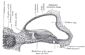
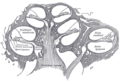
The tympanic duct is located in the cochlea, which is a spiral-shaped organ within the osseous labyrinth of the inner ear. It is situated below the cochlear duct (scala media) and is separated from it by the basilar membrane. The tympanic duct is filled with a fluid called perilymph, which is similar in composition to cerebrospinal fluid.

Function[edit]
The primary function of the tympanic duct is to transmit sound vibrations. When sound waves enter the ear, they cause the tympanic membrane (eardrum) to vibrate. These vibrations are then transmitted through the ossicles of the middle ear to the oval window, which is the entrance to the cochlea. The movement of the oval window creates waves in the perilymph of the vestibular duct (scala vestibuli), which then travel through the helicotrema to the tympanic duct. The waves in the perilymph of the tympanic duct cause the basilar membrane to move, stimulating the hair cells in the cochlear duct and ultimately leading to the perception of sound.
Clinical significance[edit]
Disorders affecting the tympanic duct can lead to hearing loss or tinnitus. Conditions such as Meniere's disease or perilymph fistula can disrupt the normal function of the tympanic duct and the cochlea as a whole.
Related structures[edit]
The tympanic duct is part of the cochlea, which also includes the vestibular duct (scala vestibuli) and the cochlear duct (scala media). These three ducts are integral to the process of hearing and are filled with different fluids that facilitate the transmission of sound waves.

See also[edit]
References[edit]
- Pickles, J. O. (2012). An Introduction to the Physiology of Hearing. Brill.
- Purves, D., Augustine, G. J., Fitzpatrick, D., et al. (2001). Neuroscience. Sinauer Associates.
Related pages[edit]
Tympanic_duct[edit]
-
 Tympanic duct
 -
 Tympanic duct


Medical Disclaimer: WikiMD is for informational purposes only and is not a substitute for professional medical advice. Content may be inaccurate or outdated and should not be used for diagnosis or treatment. Always consult your healthcare provider for medical decisions. Verify information with trusted sources such as CDC.gov and NIH.gov. By using this site, you agree that WikiMD is not liable for any outcomes related to its content. See full disclaimer.
Credits:Most images are courtesy of Wikimedia commons, and templates, categories Wikipedia, licensed under CC BY SA or similar.
Translate page: - East Asian
中文,
日本,
한국어,
South Asian
हिन्दी,
தமிழ்,
తెలుగు,
Urdu,
ಕನ್ನಡ,
Southeast Asian
Indonesian,
Vietnamese,
Thai,
မြန်မာဘာသာ,
বাংলা
European
español,
Deutsch,
français,
Greek,
português do Brasil,
polski,
română,
русский,
Nederlands,
norsk,
svenska,
suomi,
Italian
Middle Eastern & African
عربى,
Turkish,
Persian,
Hebrew,
Afrikaans,
isiZulu,
Kiswahili,
Other
Bulgarian,
Hungarian,
Czech,
Swedish,
മലയാളം,
मराठी,
ਪੰਜਾਬੀ,
ગુજરાતી,
Portuguese,
Ukrainian
