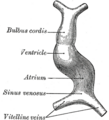
Vitelline veins
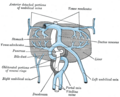
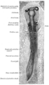
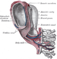
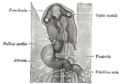
Vitelline veins are a pair of veins that drain blood from the yolk sac to the sinus venosus in the developing embryo. They are part of the cardiovascular system and play a crucial role in the formation of the portal vein and inferior vena cava.
Etymology[edit]
The term "vitelline" is derived from the Latin word "vitellus" which means "yolk". This is a reference to the veins' connection to the yolk sac in the embryo.
Development[edit]
The vitelline veins develop during the third week of embryogenesis. They initially form as a pair of symmetrical structures that drain the yolk sac into the sinus venosus. As development progresses, the veins undergo a series of transformations that result in the formation of the portal vein and the inferior vena cava.
Function[edit]
The primary function of the vitelline veins is to transport blood from the yolk sac to the heart. The yolk sac is a temporary structure that provides nutrients to the developing embryo before the placenta is fully formed. The blood carried by the vitelline veins is rich in nutrients and oxygen, which are essential for the growth and development of the embryo.
Clinical significance[edit]
Abnormalities in the development of the vitelline veins can lead to a variety of congenital heart defects. These can include portal vein thrombosis, portal hypertension, and budd-chiari syndrome. These conditions can have serious health implications and may require medical intervention.
See also[edit]
References[edit]

This WikiMD article can only be edited by registered and verified editors. You can log in or register.
-
 Vitelline veins
 -
Embryological development of the human venous system
-
 Vitelline veins
 -
 Vitelline veins
 -
 Vitelline veins
 -
 Vitelline veins
 -
 Vitelline veins


Medical Disclaimer: WikiMD is for informational purposes only and is not a substitute for professional medical advice. Content may be inaccurate or outdated and should not be used for diagnosis or treatment. Always consult your healthcare provider for medical decisions. Verify information with trusted sources such as CDC.gov and NIH.gov. By using this site, you agree that WikiMD is not liable for any outcomes related to its content. See full disclaimer.
Credits:Most images are courtesy of Wikimedia commons, and templates, categories Wikipedia, licensed under CC BY SA or similar.
Translate page: - East Asian
中文,
日本,
한국어,
South Asian
हिन्दी,
தமிழ்,
తెలుగు,
Urdu,
ಕನ್ನಡ,
Southeast Asian
Indonesian,
Vietnamese,
Thai,
မြန်မာဘာသာ,
বাংলা
European
español,
Deutsch,
français,
Greek,
português do Brasil,
polski,
română,
русский,
Nederlands,
norsk,
svenska,
suomi,
Italian
Middle Eastern & African
عربى,
Turkish,
Persian,
Hebrew,
Afrikaans,
isiZulu,
Kiswahili,
Other
Bulgarian,
Hungarian,
Czech,
Swedish,
മലയാളം,
मराठी,
ਪੰਜਾਬੀ,
ગુજરાતી,
Portuguese,
Ukrainian
