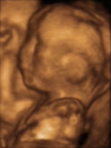
3D ultrasound
3D Ultrasound[edit | edit source]
3D ultrasound is a medical ultrasound technique, often used in obstetric ultrasonography, providing three-dimensional images of the fetus. Unlike traditional 2D ultrasound, which provides flat, two-dimensional images, 3D ultrasound offers a more detailed view, allowing for better visualization of the fetus's external features.
History[edit | edit source]
The development of 3D ultrasound technology began in the late 20th century, building upon the advancements in ultrasound imaging technology. The first 3D ultrasound images were produced in the 1980s, but it wasn't until the 1990s that the technology became more widely available in clinical practice.
Technique[edit | edit source]
3D ultrasound imaging is achieved by acquiring multiple 2D images at different angles and then reconstructing them into a three-dimensional image using specialized software. This process is known as volume rendering. The resulting images can be rotated and viewed from different angles, providing a comprehensive view of the fetus.
Applications[edit | edit source]
3D ultrasound is primarily used in prenatal diagnosis to assess fetal development and detect any abnormalities. It is particularly useful for:
- Evaluating fetal anatomy and detecting congenital anomalies.
- Assessing the development of the fetal face, limbs, and other external structures.
- Providing parents with a more realistic view of their unborn child.
Advantages[edit | edit source]
The advantages of 3D ultrasound over traditional 2D ultrasound include:
- Enhanced visualization of fetal structures, which can improve diagnostic accuracy.
- The ability to view the fetus from multiple angles, aiding in the assessment of complex anatomical structures.
- Improved parental bonding, as the realistic images can enhance the emotional connection between parents and their unborn child.
Limitations[edit | edit source]
Despite its advantages, 3D ultrasound has some limitations:
- It is more expensive and less widely available than 2D ultrasound.
- The quality of the images can be affected by factors such as fetal position, maternal obesity, and the amount of amniotic fluid.
- It requires specialized equipment and trained personnel to perform and interpret the images.
Related pages[edit | edit source]
Transform your life with W8MD's budget GLP1 injections from $125
W8MD offers a medical weight loss program NYC and a clinic to lose weight in Philadelphia. Our W8MD's physician supervised medical weight loss centers in NYC provides expert medical guidance, and offers telemedicine options for convenience.
Why choose W8MD?
- Comprehensive care with FDA-approved weight loss medications including:
- loss injections in NYC both generic and brand names:
- weight loss medications including Phentermine, Qsymia, Diethylpropion etc.
- Accept most insurances for visits or discounted self pay cost.
- Generic weight loss injections starting from just $125.00 for the starting dose
- In person weight loss NYC and telemedicine medical weight loss options in New York city available
- Budget GLP1 weight loss injections in NYC starting from $125.00 biweekly with insurance!
Book Your Appointment
Start your NYC weight loss journey today at our NYC medical weight loss, and Philadelphia medical weight loss Call (718)946-5500 for NY and 215 676 2334 for PA
Search WikiMD
Ad.Tired of being Overweight? Try W8MD's NYC physician weight loss.
Semaglutide (Ozempic / Wegovy and Tirzepatide (Mounjaro / Zepbound) available. Call 718 946 5500.
Advertise on WikiMD
|
WikiMD's Wellness Encyclopedia |
| Let Food Be Thy Medicine Medicine Thy Food - Hippocrates |
Translate this page: - East Asian
中文,
日本,
한국어,
South Asian
हिन्दी,
தமிழ்,
తెలుగు,
Urdu,
ಕನ್ನಡ,
Southeast Asian
Indonesian,
Vietnamese,
Thai,
မြန်မာဘာသာ,
বাংলা
European
español,
Deutsch,
français,
Greek,
português do Brasil,
polski,
română,
русский,
Nederlands,
norsk,
svenska,
suomi,
Italian
Middle Eastern & African
عربى,
Turkish,
Persian,
Hebrew,
Afrikaans,
isiZulu,
Kiswahili,
Other
Bulgarian,
Hungarian,
Czech,
Swedish,
മലയാളം,
मराठी,
ਪੰਜਾਬੀ,
ગુજરાતી,
Portuguese,
Ukrainian
Medical Disclaimer: WikiMD is not a substitute for professional medical advice. The information on WikiMD is provided as an information resource only, may be incorrect, outdated or misleading, and is not to be used or relied on for any diagnostic or treatment purposes. Please consult your health care provider before making any healthcare decisions or for guidance about a specific medical condition. WikiMD expressly disclaims responsibility, and shall have no liability, for any damages, loss, injury, or liability whatsoever suffered as a result of your reliance on the information contained in this site. By visiting this site you agree to the foregoing terms and conditions, which may from time to time be changed or supplemented by WikiMD. If you do not agree to the foregoing terms and conditions, you should not enter or use this site. See full disclaimer.
Credits:Most images are courtesy of Wikimedia commons, and templates, categories Wikipedia, licensed under CC BY SA or similar.
Contributors: Prab R. Tumpati, MD


