Anterior external arcuate fibers
Anterior External Arcuate Fibers[edit]
The anterior external arcuate fibers are a group of nerve fibers located in the brain. They are part of the white matter tracts that connect different regions of the brain, specifically the frontal and temporal lobes. In this article, we will explore the anatomy, function, and clinical significance of the anterior external arcuate fibers.
Anatomy[edit]
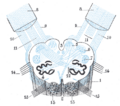
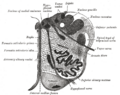
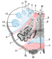
The anterior external arcuate fibers are located in the cerebral hemisphere, specifically in the white matter of the brain. They are part of the superior longitudinal fasciculus (SLF), which is a bundle of nerve fibers that connects the frontal, parietal, and temporal lobes.
The SLF can be divided into three major components: SLF I, SLF II, and SLF III. The anterior external arcuate fibers are a part of SLF III. They originate from the frontal lobe, specifically the prefrontal cortex, and extend towards the temporal lobe.
Function[edit]
The exact function of the anterior external arcuate fibers is not fully understood. However, studies suggest that they play a role in language processing and auditory integration. They are believed to be involved in the integration of auditory information with other cognitive processes, such as attention and memory.
The anterior external arcuate fibers are also thought to be involved in the control of eye movements. They connect the frontal eye fields, which are responsible for voluntary eye movements, with the temporal lobe, which is involved in visual processing.
Clinical Significance[edit]
Damage or disruption to the anterior external arcuate fibers can result in various neurological conditions. For example, lesions in this area have been associated with language disorders, such as aphasia. Patients with damage to these fibers may have difficulty understanding or producing language.
Additionally, abnormalities in the anterior external arcuate fibers have been observed in individuals with schizophrenia. This suggests a potential role of these fibers in the pathophysiology of the disorder.
Conclusion[edit]
The anterior external arcuate fibers are an important component of the white matter tracts in the brain. They connect the frontal and temporal lobes and are involved in language processing, auditory integration, and eye movement control. Understanding the anatomy and function of these fibers can provide valuable insights into various neurological conditions and contribute to the development of targeted therapies.
See Also[edit]
-
 Anterior external arcuate fibers
 -
 Anterior external arcuate fibers
 -
 Anterior external arcuate fibers


Medical Disclaimer: WikiMD is for informational purposes only and is not a substitute for professional medical advice. Content may be inaccurate or outdated and should not be used for diagnosis or treatment. Always consult your healthcare provider for medical decisions. Verify information with trusted sources such as CDC.gov and NIH.gov. By using this site, you agree that WikiMD is not liable for any outcomes related to its content. See full disclaimer.
Credits:Most images are courtesy of Wikimedia commons, and templates, categories Wikipedia, licensed under CC BY SA or similar.
Translate page: - East Asian
中文,
日本,
한국어,
South Asian
हिन्दी,
தமிழ்,
తెలుగు,
Urdu,
ಕನ್ನಡ,
Southeast Asian
Indonesian,
Vietnamese,
Thai,
မြန်မာဘာသာ,
বাংলা
European
español,
Deutsch,
français,
Greek,
português do Brasil,
polski,
română,
русский,
Nederlands,
norsk,
svenska,
suomi,
Italian
Middle Eastern & African
عربى,
Turkish,
Persian,
Hebrew,
Afrikaans,
isiZulu,
Kiswahili,
Other
Bulgarian,
Hungarian,
Czech,
Swedish,
മലയാളം,
मराठी,
ਪੰਜਾਬੀ,
ગુજરાતી,
Portuguese,
Ukrainian
