Automated tissue image analysis
Automated Tissue Image Analysis is a branch of digital pathology that involves the use of computer technology to assist in the interpretation and analysis of tissue samples obtained during biopsy or surgery. This technology leverages machine learning and image processing techniques to enhance the accuracy and efficiency of pathological assessment, which is crucial in the diagnosis and treatment planning for various diseases, including cancer.
Overview[edit]
Automated tissue image analysis systems utilize advanced algorithms to analyze high-resolution images of tissue samples. These systems are designed to identify and quantify morphological features such as cell size, shape, and organization, as well as to detect the presence of specific biomarkers that can indicate disease. By automating the analysis process, these systems can reduce the subjectivity and variability associated with manual microscopy, leading to more consistent and reliable results.
Applications[edit]
The primary application of automated tissue image analysis is in the field of oncology, where it is used to improve the accuracy of cancer diagnosis and to assess tumor grade and stage. This technology can also be applied in the research setting, enabling scientists to perform large-scale analysis of tissue samples for the study of disease mechanisms and the development of new therapeutic strategies.
Advantages[edit]
- Increased Efficiency: Automated analysis can process large volumes of tissue images much faster than manual methods, significantly reducing the time required for diagnosis.
- Enhanced Accuracy: By minimizing human error and variability, automated systems can provide more consistent and objective assessments of tissue samples.
- Quantitative Analysis: Automated systems can provide quantitative measurements of tissue features, offering more detailed insights into disease pathology.
Challenges[edit]
Despite its benefits, the implementation of automated tissue image analysis faces several challenges. These include the need for high-quality imaging equipment, the complexity of developing accurate and robust algorithms, and the requirement for extensive validation to ensure reliability across different types of tissue samples. Additionally, there is an ongoing need for collaboration between pathologists and engineers to refine these systems and integrate them effectively into clinical practice.
Future Directions[edit]
The field of automated tissue image analysis is rapidly evolving, with ongoing research focused on improving the accuracy and versatility of these systems. Future developments may include the integration of artificial intelligence and deep learning techniques to further enhance diagnostic capabilities, as well as the expansion of applications beyond oncology to other areas of pathology.
This medical article is a stub. You can help WikiMD by expanding the page. |
Automated tissue image analysis[edit]
-
 Microscope with stained slide
 -
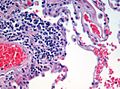 Emphysema H and E


Medical Disclaimer: WikiMD is for informational purposes only and is not a substitute for professional medical advice. Content may be inaccurate or outdated and should not be used for diagnosis or treatment. Always consult your healthcare provider for medical decisions. Verify information with trusted sources such as CDC.gov and NIH.gov. By using this site, you agree that WikiMD is not liable for any outcomes related to its content. See full disclaimer.
Credits:Most images are courtesy of Wikimedia commons, and templates, categories Wikipedia, licensed under CC BY SA or similar.
Translate page: - East Asian
中文,
日本,
한국어,
South Asian
हिन्दी,
தமிழ்,
తెలుగు,
Urdu,
ಕನ್ನಡ,
Southeast Asian
Indonesian,
Vietnamese,
Thai,
မြန်မာဘာသာ,
বাংলা
European
español,
Deutsch,
français,
Greek,
português do Brasil,
polski,
română,
русский,
Nederlands,
norsk,
svenska,
suomi,
Italian
Middle Eastern & African
عربى,
Turkish,
Persian,
Hebrew,
Afrikaans,
isiZulu,
Kiswahili,
Other
Bulgarian,
Hungarian,
Czech,
Swedish,
മലയാളം,
मराठी,
ਪੰਜਾਬੀ,
ગુજરાતી,
Portuguese,
Ukrainian
