Cardiac fibrosis
Cardiac fibrosis is a pathological condition characterized by the excessive accumulation of extracellular matrix proteins, particularly collagen, in the heart muscle. This process leads to the stiffening of the heart tissues, impairing the organ's ability to contract and relax efficiently, which can significantly affect heart function and lead to various cardiovascular diseases, including heart failure, cardiomyopathy, and arrhythmia.
Etiology[edit]
Cardiac fibrosis can be caused by several factors, including long-standing hypertension, myocardial infarction (heart attack), cardiomyopathies (diseases of the heart muscle), and exposure to certain toxins. Chronic high blood pressure forces the heart to work harder than normal, leading to structural changes in the heart muscle and the eventual development of fibrosis. Similarly, after a myocardial infarction, the healing process involves the replacement of dead cardiac muscle cells with fibrotic tissue, which can disrupt the normal architecture and function of the heart.
Pathophysiology[edit]
The pathophysiology of cardiac fibrosis involves complex signaling pathways that lead to the activation of fibroblasts, the cells responsible for collagen production. Under pathological conditions, fibroblasts transform into myofibroblasts, which have a higher capacity for collagen synthesis. Key mediators of this process include transforming growth factor-beta (TGF-β), angiotensin II, and platelet-derived growth factor (PDGF), among others. The excessive deposition of collagen and other extracellular matrix components results in the thickening and stiffening of the heart walls, reducing the heart's elasticity and contractile function.
Clinical Manifestations[edit]
The clinical manifestations of cardiac fibrosis can vary widely depending on the extent and location of fibrosis. Common symptoms include shortness of breath, fatigue, reduced exercise capacity, and in more severe cases, heart failure. Cardiac fibrosis can also lead to arrhythmias due to the disruption of normal electrical conduction pathways in the heart.
Diagnosis[edit]
The diagnosis of cardiac fibrosis typically involves a combination of patient history, physical examination, and diagnostic tests. Imaging modalities such as echocardiography and cardiac magnetic resonance imaging (MRI) can be used to assess the structure and function of the heart and detect signs of fibrosis. In some cases, a cardiac biopsy may be performed to obtain a definitive diagnosis, though this is less common due to its invasive nature.
Treatment[edit]
Treatment of cardiac fibrosis focuses on managing the underlying cause and preventing further progression of the disease. This may include the use of medications such as angiotensin-converting enzyme inhibitors (ACE inhibitors), angiotensin receptor blockers (ARBs), and beta-blockers to control blood pressure and reduce cardiac workload. In cases where arrhythmias are present, anti-arrhythmic drugs or devices such as pacemakers may be necessary. Currently, there are no specific treatments that can reverse cardiac fibrosis, making early detection and management of risk factors critical.
Research Directions[edit]
Research into cardiac fibrosis is ongoing, with studies focusing on understanding the molecular mechanisms underlying the condition and developing targeted therapies. Novel approaches such as the use of microRNAs and stem cell therapy are being explored as potential treatments to modulate fibroblast activity and promote the regeneration of healthy cardiac tissue.
-
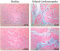 Histopathology of interstitial fibrosis in dilated cardiomyopathy
 -
Histopathology of interstitial fibrosis of chronic ischemic heart disease
-
Subepicardial fibrosis
-
 Histopathology of dense fibrous scar replacing myocyte loss in myocardial infarction


Medical Disclaimer: WikiMD is for informational purposes only and is not a substitute for professional medical advice. Content may be inaccurate or outdated and should not be used for diagnosis or treatment. Always consult your healthcare provider for medical decisions. Verify information with trusted sources such as CDC.gov and NIH.gov. By using this site, you agree that WikiMD is not liable for any outcomes related to its content. See full disclaimer.
Credits:Most images are courtesy of Wikimedia commons, and templates, categories Wikipedia, licensed under CC BY SA or similar.
Translate page: - East Asian
中文,
日本,
한국어,
South Asian
हिन्दी,
தமிழ்,
తెలుగు,
Urdu,
ಕನ್ನಡ,
Southeast Asian
Indonesian,
Vietnamese,
Thai,
မြန်မာဘာသာ,
বাংলা
European
español,
Deutsch,
français,
Greek,
português do Brasil,
polski,
română,
русский,
Nederlands,
norsk,
svenska,
suomi,
Italian
Middle Eastern & African
عربى,
Turkish,
Persian,
Hebrew,
Afrikaans,
isiZulu,
Kiswahili,
Other
Bulgarian,
Hungarian,
Czech,
Swedish,
മലയാളം,
मराठी,
ਪੰਜਾਬੀ,
ગુજરાતી,
Portuguese,
Ukrainian
