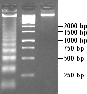
DNA laddering
DNA Laddering[edit | edit source]
DNA laddering is a distinctive pattern of DNA fragments that is often observed during apoptosis, a form of programmed cell death. This pattern is characterized by the appearance of DNA fragments that are multiples of approximately 180-200 base pairs in length, which can be visualized using agarose gel electrophoresis.
Mechanism[edit | edit source]
DNA laddering occurs as a result of the activation of specific endonucleases during apoptosis. These enzymes cleave the chromosomal DNA at internucleosomal regions, leading to the formation of DNA fragments of regular sizes. The regularity of these fragments is due to the periodicity of nucleosomes, which are the fundamental units of chromatin structure, consisting of DNA wrapped around histone proteins.
Detection[edit | edit source]
The detection of DNA laddering is a common method used to confirm the occurrence of apoptosis in cell samples. During the process, cells are lysed to release their DNA, which is then subjected to agarose gel electrophoresis. The DNA fragments migrate through the gel, and when stained with a DNA-binding dye such as ethidium bromide, they appear as a series of bands resembling a "ladder".
Significance[edit | edit source]
DNA laddering is considered a hallmark of apoptosis and is used in research to distinguish apoptotic cell death from necrosis, which typically results in a smear of DNA rather than discrete bands. This method is widely used in molecular biology and biochemistry to study cell death mechanisms and to evaluate the effects of various treatments on cell viability.
Related Pages[edit | edit source]
Search WikiMD
Ad.Tired of being Overweight? Try W8MD's physician weight loss program.
Semaglutide (Ozempic / Wegovy and Tirzepatide (Mounjaro / Zepbound) available.
Advertise on WikiMD
|
WikiMD's Wellness Encyclopedia |
| Let Food Be Thy Medicine Medicine Thy Food - Hippocrates |
Translate this page: - East Asian
中文,
日本,
한국어,
South Asian
हिन्दी,
தமிழ்,
తెలుగు,
Urdu,
ಕನ್ನಡ,
Southeast Asian
Indonesian,
Vietnamese,
Thai,
မြန်မာဘာသာ,
বাংলা
European
español,
Deutsch,
français,
Greek,
português do Brasil,
polski,
română,
русский,
Nederlands,
norsk,
svenska,
suomi,
Italian
Middle Eastern & African
عربى,
Turkish,
Persian,
Hebrew,
Afrikaans,
isiZulu,
Kiswahili,
Other
Bulgarian,
Hungarian,
Czech,
Swedish,
മലയാളം,
मराठी,
ਪੰਜਾਬੀ,
ગુજરાતી,
Portuguese,
Ukrainian
Medical Disclaimer: WikiMD is not a substitute for professional medical advice. The information on WikiMD is provided as an information resource only, may be incorrect, outdated or misleading, and is not to be used or relied on for any diagnostic or treatment purposes. Please consult your health care provider before making any healthcare decisions or for guidance about a specific medical condition. WikiMD expressly disclaims responsibility, and shall have no liability, for any damages, loss, injury, or liability whatsoever suffered as a result of your reliance on the information contained in this site. By visiting this site you agree to the foregoing terms and conditions, which may from time to time be changed or supplemented by WikiMD. If you do not agree to the foregoing terms and conditions, you should not enter or use this site. See full disclaimer.
Credits:Most images are courtesy of Wikimedia commons, and templates, categories Wikipedia, licensed under CC BY SA or similar.
Contributors: Prab R. Tumpati, MD

