Doppler echocardiography
Doppler echocardiography is a diagnostic procedure that uses ultrasound waves to produce images of the heart and to measure the speed and direction of blood flow within the heart chambers and blood vessels. This technique is an integral part of echocardiography, combining traditional ultrasound imaging with the Doppler effect to evaluate both the structure and function of the heart. Doppler echocardiography is particularly useful in diagnosing various heart conditions, including heart valve diseases, congenital heart defects, and heart failure, among others.
Principles of Doppler Echocardiography[edit]
The underlying principle of Doppler echocardiography is based on the Doppler effect, which describes the change in frequency or wavelength of a wave in relation to an observer moving relative to the source of the wave. In the context of echocardiography, this principle is applied to sound waves that are emitted by a transducer, reflect off blood cells moving within the heart and blood vessels, and then return to the transducer. The change in frequency of the returning sound waves is directly proportional to the speed and direction of the blood flow, allowing for detailed assessment of blood flow patterns and velocities.
Types of Doppler Echocardiography[edit]
There are several types of Doppler echocardiography, each with specific applications:
- Continuous Wave Doppler: This method captures high-velocity blood flow, such as that seen in severe valvular stenosis or regurgitation. It uses two crystals in the transducer; one continuously transmits sound waves while the other continuously receives them.
- Pulsed Wave Doppler: Pulsed wave Doppler allows for the measurement of blood flow velocities at specific locations within the heart. This is particularly useful for evaluating blood flow across heart valves.
- Color Flow Doppler: This technique adds a color overlay to traditional 2D echocardiogram images, with different colors representing the direction of blood flow. This helps in visualizing the flow patterns within the heart chambers and through the valves.
- Tissue Doppler Imaging (TDI): TDI measures the velocity of the heart muscle (myocardium) movements, aiding in the assessment of diastolic function and cardiac synchrony.
Clinical Applications[edit]
Doppler echocardiography is used in the diagnosis and management of a wide range of cardiac conditions:
- Evaluating the presence and severity of valvular heart disease, including stenosis and regurgitation.
- Assessing left and right ventricular systolic and diastolic function.
- Detecting congenital heart defects such as atrial septal defect (ASD) and ventricular septal defect (VSD).
- Guiding the treatment of patients with heart failure by assessing cardiac output and filling pressures.
- Monitoring the status of heart valve prostheses.
- Evaluating the effectiveness of medical or surgical treatments over time.
Advantages and Limitations[edit]
Doppler echocardiography offers several advantages, including its non-invasive nature, absence of ionizing radiation, and its ability to provide real-time dynamic images of the heart and blood flow. However, its effectiveness can be limited by factors such as patient body habitus, lung interference, and the skill and experience of the operator.
Conclusion[edit]
Doppler echocardiography is a versatile and invaluable tool in the field of cardiology, providing critical information on heart structure, function, and blood flow. Its non-invasive nature and comprehensive diagnostic capabilities make it an essential component of cardiac assessment and management.
This medical article is a stub. You can help WikiMD by expanding the page. |
-
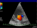 Doppler echocardiography of mitral valve
 -
 Interpolation to find peak
 -
 Lateral phase cross-correlation


Medical Disclaimer: WikiMD is for informational purposes only and is not a substitute for professional medical advice. Content may be inaccurate or outdated and should not be used for diagnosis or treatment. Always consult your healthcare provider for medical decisions. Verify information with trusted sources such as CDC.gov and NIH.gov. By using this site, you agree that WikiMD is not liable for any outcomes related to its content. See full disclaimer.
Credits:Most images are courtesy of Wikimedia commons, and templates, categories Wikipedia, licensed under CC BY SA or similar.
Translate page: - East Asian
中文,
日本,
한국어,
South Asian
हिन्दी,
தமிழ்,
తెలుగు,
Urdu,
ಕನ್ನಡ,
Southeast Asian
Indonesian,
Vietnamese,
Thai,
မြန်မာဘာသာ,
বাংলা
European
español,
Deutsch,
français,
Greek,
português do Brasil,
polski,
română,
русский,
Nederlands,
norsk,
svenska,
suomi,
Italian
Middle Eastern & African
عربى,
Turkish,
Persian,
Hebrew,
Afrikaans,
isiZulu,
Kiswahili,
Other
Bulgarian,
Hungarian,
Czech,
Swedish,
മലയാളം,
मराठी,
ਪੰਜਾਬੀ,
ગુજરાતી,
Portuguese,
Ukrainian
