Fluorescence spectroscopy
Fluorescence spectroscopy, also known as fluorometry or spectrofluorometry, is a type of electromagnetic spectroscopy which analyzes fluorescence from a sample. It involves using a beam of light, usually ultraviolet, to excite the electrons in molecules of certain compounds and cause them to emit light of a lower energy, typically, but not exclusively, visible light. This technique has wide applications in various fields such as chemistry, biochemistry, medical diagnostics, and molecular biology, providing qualitative and quantitative data about the structure and composition of substances.
Principles[edit]
The fundamental principle of fluorescence spectroscopy is based on the absorption and subsequent emission of light by a substance. When molecules absorb light at a specific wavelength, they transition from a ground state to an excited state. As they return to their ground state, they emit photons at a longer wavelength than the absorbed light. This phenomenon is known as fluorescence. The difference in energy between the absorbed and emitted light is characteristic of the specific material and is used for identification and quantification.
Instrumentation[edit]
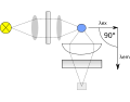
A basic fluorescence spectrometer consists of a light source, typically a xenon or mercury lamp, to provide the excitation light. A monochromator selects the wavelength of the excitation light that is directed towards the sample. The emitted light is then collected at a 90-degree angle to the incident light to minimize the detection of reflected excitation light. Another monochromator selects the wavelength of the emitted light, which is detected by a photodetector, such as a photomultiplier tube (PMT) or a charge-coupled device (CCD).
Applications[edit]
Fluorescence spectroscopy has diverse applications across various scientific disciplines:
- In biochemistry and molecular biology, it is used to study the structure, dynamics, and interactions of biomolecules. For example, the technique can be used to monitor the folding and unfolding of proteins.
- In medical diagnostics, fluorescence spectroscopy can detect and quantify biomarkers in tissue samples, aiding in the diagnosis of diseases such as cancer.
- In environmental science, it is employed to detect pollutants in water and air samples.
- In material science, it helps in the characterization of materials, including the study of semiconductor nanocrystals and organic light-emitting diodes (OLEDs).
Advantages and Limitations[edit]
The main advantage of fluorescence spectroscopy is its high sensitivity and specificity, allowing for the detection of low concentrations of substances. It also provides rich information about the molecular environment of the fluorescent species. However, the technique has limitations, including photobleaching (the irreversible destruction of a fluorophore), fluorescence quenching, and the requirement for the sample to be fluorescent or to be labeled with a fluorescent tag.
See Also[edit]
This biochemistry article is a stub. You can help WikiMD by expanding the page. |
This medical article is a stub. You can help WikiMD by expanding the page. |
-
 Fluorescence spectroscopy
 -
 Fluorimeter used in fluorescence spectroscopy
 -
 OpenFluor R Script Export
 -
 OpenFluor Models Report


Medical Disclaimer: WikiMD is for informational purposes only and is not a substitute for professional medical advice. Content may be inaccurate or outdated and should not be used for diagnosis or treatment. Always consult your healthcare provider for medical decisions. Verify information with trusted sources such as CDC.gov and NIH.gov. By using this site, you agree that WikiMD is not liable for any outcomes related to its content. See full disclaimer.
Credits:Most images are courtesy of Wikimedia commons, and templates, categories Wikipedia, licensed under CC BY SA or similar.
Translate page: - East Asian
中文,
日本,
한국어,
South Asian
हिन्दी,
தமிழ்,
తెలుగు,
Urdu,
ಕನ್ನಡ,
Southeast Asian
Indonesian,
Vietnamese,
Thai,
မြန်မာဘာသာ,
বাংলা
European
español,
Deutsch,
français,
Greek,
português do Brasil,
polski,
română,
русский,
Nederlands,
norsk,
svenska,
suomi,
Italian
Middle Eastern & African
عربى,
Turkish,
Persian,
Hebrew,
Afrikaans,
isiZulu,
Kiswahili,
Other
Bulgarian,
Hungarian,
Czech,
Swedish,
മലയാളം,
मराठी,
ਪੰਜਾਬੀ,
ગુજરાતી,
Portuguese,
Ukrainian
