Fundus photography
Fundus Photography is a specialized form of medical imaging that provides a detailed view of the retina, optic disc, macula, and the posterior pole of the eye. It is a crucial tool in the diagnosis and management of various eye diseases and conditions, including diabetic retinopathy, macular degeneration, and glaucoma.
History[edit]
The concept of fundus photography was first introduced in the late 19th century, with the development of the first ophthalmoscope by Hermann von Helmholtz. The technology has since evolved, with modern fundus cameras capable of capturing high-resolution images of the eye's interior.
Technique[edit]
Fundus photography involves the use of a specialized low-power microscope with an attached camera known as a fundus camera. The patient's eye is dilated using mydriatic eye drops to allow a clear view of the fundus. The camera then captures images of the fundus, which can be stored digitally for further analysis and comparison over time.
Applications[edit]
Fundus photography is used in a variety of clinical settings, including ophthalmology, optometry, and neurology. It is particularly useful in the diagnosis and monitoring of diseases that affect the retina and other structures at the back of the eye.
Limitations[edit]
While fundus photography provides valuable information about the eye's interior, it does have some limitations. It only captures a small portion of the retina at a time, and it may not provide a clear view of the peripheral retina. Additionally, it may not be suitable for patients with certain eye conditions, such as cataracts, that can obstruct the view of the fundus.
Future Developments[edit]
Advancements in technology are continually improving the capabilities of fundus photography. Developments in digital imaging, artificial intelligence, and telemedicine are expected to enhance the diagnostic accuracy and accessibility of this important tool in eye care.
This medical article is a stub. You can help WikiMD by expanding the page. |
This ophthalmology article is a stub. You can help WikiMD by expanding the page. |
-
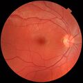
 Fundus photograph of normal left eye
 -
 Fundus photograph of normal right eye
 -
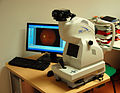 Fundus camera
 -
 Retina camera controls
 -
 Fundus photo showing focal laser surgery for diabetic retinopathy


Medical Disclaimer: WikiMD is for informational purposes only and is not a substitute for professional medical advice. Content may be inaccurate or outdated and should not be used for diagnosis or treatment. Always consult your healthcare provider for medical decisions. Verify information with trusted sources such as CDC.gov and NIH.gov. By using this site, you agree that WikiMD is not liable for any outcomes related to its content. See full disclaimer.
Credits:Most images are courtesy of Wikimedia commons, and templates, categories Wikipedia, licensed under CC BY SA or similar.
Translate page: - East Asian
中文,
日本,
한국어,
South Asian
हिन्दी,
தமிழ்,
తెలుగు,
Urdu,
ಕನ್ನಡ,
Southeast Asian
Indonesian,
Vietnamese,
Thai,
မြန်မာဘာသာ,
বাংলা
European
español,
Deutsch,
français,
Greek,
português do Brasil,
polski,
română,
русский,
Nederlands,
norsk,
svenska,
suomi,
Italian
Middle Eastern & African
عربى,
Turkish,
Persian,
Hebrew,
Afrikaans,
isiZulu,
Kiswahili,
Other
Bulgarian,
Hungarian,
Czech,
Swedish,
മലയാളം,
मराठी,
ਪੰਜਾਬੀ,
ગુજરાતી,
Portuguese,
Ukrainian
