Phase-contrast microscopy
Phase-contrast microscopy is a microscopy technique that converts phase shifts in light passing through a transparent specimen to brightness changes in the image. Developed by Dutch physicist Frits Zernike in the 1930s, for which he was awarded the Nobel Prize in Physics in 1953, this technique became an essential tool in biological and medical research, allowing for the visualization of cellular structures and organelles without the need for staining or fixing.
Principle[edit]
The principle of phase-contrast microscopy lies in the manipulation of the optical path of light. As light waves pass through a specimen, they undergo phase shifts due to variations in the refractive index of different parts of the specimen. These phase shifts, however, are not discernible to the human eye. Phase-contrast microscopy employs a special device called a phase plate to convert these phase shifts into variations in light intensity, making the transparent details of the specimen visible.
Components[edit]
The key components of a phase-contrast microscope include:
- Condenser annulus: A ring-shaped aperture located in the condenser that produces a cone of light focused on the specimen.
- Phase plate: Located in the objective lens, it introduces a phase shift to the undeviated light that has passed directly through the specimen, enhancing the contrast between different parts of the specimen.
- Objective lens: Collects the light from the specimen and forms the image.
Applications[edit]
Phase-contrast microscopy is widely used in various fields of biological and medical research. Its applications include:
- Observing living cells and tissues, allowing researchers to study cellular processes in real time.
- Examining microorganisms, such as bacteria and protozoa, without the need for staining.
- Analyzing sperm motility in medical diagnostics.
- Investigating the structure of fibers and polymers in materials science.
Advantages and Limitations[edit]
Advantages:
- Allows for the observation of living cells without the need for staining or fixation.
- Enhances the contrast of transparent specimens, making it easier to study their internal structures.
Limitations:
- Phase halos, a form of optical artifact, can occur around the edges of high-contrast structures.
- Limited to specimens that are thin enough to allow for phase shifts within the light path.
See Also[edit]
References[edit]
| Optical microscopy | ||||||
|---|---|---|---|---|---|---|
|
This Microscopy-related article is a stub. You can help WikiMD by expanding the page. |
-
Phase contrast microscope
-
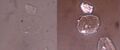 Brightfield phase contrast cell image
 -
Working principle of phase contrast microscopy
-
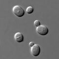 S cerevisiae under DIC microscopy
 -
Phase shift image of cells in 3D


Medical Disclaimer: WikiMD is for informational purposes only and is not a substitute for professional medical advice. Content may be inaccurate or outdated and should not be used for diagnosis or treatment. Always consult your healthcare provider for medical decisions. Verify information with trusted sources such as CDC.gov and NIH.gov. By using this site, you agree that WikiMD is not liable for any outcomes related to its content. See full disclaimer.
Credits:Most images are courtesy of Wikimedia commons, and templates, categories Wikipedia, licensed under CC BY SA or similar.
Translate page: - East Asian
中文,
日本,
한국어,
South Asian
हिन्दी,
தமிழ்,
తెలుగు,
Urdu,
ಕನ್ನಡ,
Southeast Asian
Indonesian,
Vietnamese,
Thai,
မြန်မာဘာသာ,
বাংলা
European
español,
Deutsch,
français,
Greek,
português do Brasil,
polski,
română,
русский,
Nederlands,
norsk,
svenska,
suomi,
Italian
Middle Eastern & African
عربى,
Turkish,
Persian,
Hebrew,
Afrikaans,
isiZulu,
Kiswahili,
Other
Bulgarian,
Hungarian,
Czech,
Swedish,
മലയാളം,
मराठी,
ਪੰਜਾਬੀ,
ગુજરાતી,
Portuguese,
Ukrainian
