Thioproperazine
Talairach Coordinates are a set of three numbers used to indicate the location of a specific point in the brain. These coordinates are based on a brain atlas developed by Jean Talairach and Pierre Tournoux. The Talairach Coordinates system is widely used in neuroimaging studies to report the location of activation foci.
Overview[edit]
The Talairach Atlas was developed in the 1960s and 1970s by Jean Talairach and Pierre Tournoux at the Sainte-Anne Hospital Center in Paris, France. The atlas is based on the post-mortem examination of a single 60-year-old French woman's brain. The brain was cut into 2mm slices and photographed, and these images were used to create a three-dimensional grid system. This grid system is the basis of the Talairach Coordinates.
Use in Neuroimaging[edit]
In neuroimaging studies, the Talairach Coordinates are used to report the location of activation foci. The coordinates are given as a set of three numbers, representing the distance (in millimeters) from the Anterior Commissure (AC) in the left-right (x), anterior-posterior (y), and inferior-superior (z) directions. The AC is used as the origin of the coordinate system because it is a clearly visible landmark in MRI images.
Criticisms and Limitations[edit]
Despite its widespread use, the Talairach Coordinate system has been criticized for several reasons. First, it is based on the brain of a single individual, and there is considerable variability in brain anatomy between individuals. Second, the original Talairach Atlas was based on post-mortem tissue, which can differ in appearance from living tissue. Finally, the atlas is based on 2D images, which can be difficult to align with 3D MRI data.
See Also[edit]
References[edit]

This WikiMD article can only be edited by registered and verified editors. You can log in or register.
-
 Chemical structure of Thioproperazine
 -
Synthesis pathway of Thioproperazine
-
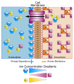 Diagram showing the basis of membrane potential


Medical Disclaimer: WikiMD is for informational purposes only and is not a substitute for professional medical advice. Content may be inaccurate or outdated and should not be used for diagnosis or treatment. Always consult your healthcare provider for medical decisions. Verify information with trusted sources such as CDC.gov and NIH.gov. By using this site, you agree that WikiMD is not liable for any outcomes related to its content. See full disclaimer.
Credits:Most images are courtesy of Wikimedia commons, and templates, categories Wikipedia, licensed under CC BY SA or similar.
Translate page: - East Asian
中文,
日本,
한국어,
South Asian
हिन्दी,
தமிழ்,
తెలుగు,
Urdu,
ಕನ್ನಡ,
Southeast Asian
Indonesian,
Vietnamese,
Thai,
မြန်မာဘာသာ,
বাংলা
European
español,
Deutsch,
français,
Greek,
português do Brasil,
polski,
română,
русский,
Nederlands,
norsk,
svenska,
suomi,
Italian
Middle Eastern & African
عربى,
Turkish,
Persian,
Hebrew,
Afrikaans,
isiZulu,
Kiswahili,
Other
Bulgarian,
Hungarian,
Czech,
Swedish,
മലയാളം,
मराठी,
ਪੰਜਾਬੀ,
ગુજરાતી,
Portuguese,
Ukrainian
