Volkmann's canal
Volkmann's canals, also known as perforating canals, are microscopic structures found in compact bone. They play a crucial role in connecting the vascular and nerve supply of the periosteum to the Haversian canals (or central canals) of the osteons.
Structure[edit]
Volkmann's canals are:
- Oriented perpendicularly or obliquely to the Haversian canals.
- Lined by endosteum, a layer of connective tissue that contains osteoblasts and osteoclasts.
- Filled with blood vessels, lymphatic vessels, and nerves that support bone health and communication.
Function[edit]
Volkmann's canals serve several important functions:
- Vascular connection: They facilitate the flow of blood between the periosteum and the Haversian canals, ensuring nutrient and oxygen delivery to osteocytes.
- Nerve transmission: They provide a pathway for nerve fibers to penetrate compact bone.
- Lymphatic drainage: They help drain lymph from the bone, maintaining tissue homeostasis.
Relationship with Haversian System[edit]
The Haversian system (or osteon) is the basic structural unit of compact bone. Volkmann's canals:
- Interconnect adjacent Haversian canals.
- Lack the concentric lamellae seen in Haversian canals.
- Provide an integrated network for vascular and neural connections across the bone.
Clinical Significance[edit]
Disruption of Volkmann's canals can occur in certain medical conditions, such as:
- Osteoporosis: Reduced bone density can impair the integrity of Volkmann's canals.
- Bone fractures: Damage to Volkmann's canals may compromise blood and nerve supply to the bone.
- Osteomyelitis: Infection within the bone can spread through these canals.
History[edit]
Volkmann's canals are named after the German physiologist Alfred Wilhelm Volkmann (1800–1877), who studied bone anatomy extensively.
See Also[edit]
References[edit]
- Gray, H. "Gray's Anatomy: The Anatomical Basis of Clinical Practice." Elsevier, 2021.
- Wheater, P.R., Burkitt, H.G., and Daniels, V.G. "Functional Histology." Churchill Livingstone, 2014.
-
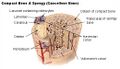 Diagram of compact and spongy bone
 -
 Bone decalcification at 40x magnification
 -
 Bone decalcification at 100x magnification


Medical Disclaimer: WikiMD is for informational purposes only and is not a substitute for professional medical advice. Content may be inaccurate or outdated and should not be used for diagnosis or treatment. Always consult your healthcare provider for medical decisions. Verify information with trusted sources such as CDC.gov and NIH.gov. By using this site, you agree that WikiMD is not liable for any outcomes related to its content. See full disclaimer.
Credits:Most images are courtesy of Wikimedia commons, and templates, categories Wikipedia, licensed under CC BY SA or similar.
Translate page: - East Asian
中文,
日本,
한국어,
South Asian
हिन्दी,
தமிழ்,
తెలుగు,
Urdu,
ಕನ್ನಡ,
Southeast Asian
Indonesian,
Vietnamese,
Thai,
မြန်မာဘာသာ,
বাংলা
European
español,
Deutsch,
français,
Greek,
português do Brasil,
polski,
română,
русский,
Nederlands,
norsk,
svenska,
suomi,
Italian
Middle Eastern & African
عربى,
Turkish,
Persian,
Hebrew,
Afrikaans,
isiZulu,
Kiswahili,
Other
Bulgarian,
Hungarian,
Czech,
Swedish,
മലയാളം,
मराठी,
ਪੰਜਾਬੀ,
ગુજરાતી,
Portuguese,
Ukrainian
