Wright's stain
Wright's stain is a histological stain that facilitates the differentiation of blood cell types. It is a commonly used stain in both hematology and the study of blood and bone marrow specimens. The stain is named after James Homer Wright, who devised the stain at the end of the 19th century. Wright's stain is a combination of eosin (a red dye) and methylene blue in methanol, which, when applied to a blood smear, yields distinct colors for different types of cells, allowing for their identification and study.
Composition and Mechanism[edit]
Wright's stain works through the principle of acid-base staining. Eosin is acidic and stains basic (or alkaline) components of the cells, such as the cytoplasm, a pink or red color. Methylene blue is basic and stains acidic components, such as nucleic acids within the nucleus, a blue or purple color. This differential staining reveals detailed structures within the cells, such as the nucleus, cytoplasm, and granules, making it easier to distinguish between different types of blood cells.
Procedure[edit]
The staining procedure involves preparing a thin film of blood on a slide, which is then fixed with methanol. The Wright's stain is applied for a specific time, followed by a rinse with a buffered water solution. The slide is then dried and can be examined under a microscope. The specific timing and concentration of the stain and buffer solution can vary, and optimal results may require adjustment based on specific laboratory conditions.
Applications[edit]
Wright's stain is extensively used in the field of hematology for performing a Complete Blood Count (CBC) and differential blood count. This staining technique allows for the identification and enumeration of different types of blood cells, including erythrocytes (red blood cells), leukocytes (white blood cells), and platelets. It is particularly useful in diagnosing blood disorders, such as anemia, leukemia, and infections, by revealing abnormalities in blood cell size, shape, and number.
Types of Cells Identified[edit]
- Erythrocytes: Appear as pink or red discs without a nucleus.
- Leukocytes:
- Neutrophils: Show a multi-lobed nucleus and light pink granules.
- Lymphocytes: Have a large, round nucleus with a thin rim of cytoplasm.
- Monocytes: Feature a large, kidney-shaped nucleus.
- Eosinophils: Contain bright red-orange granules.
- Basophils: Display dark purple-black granules.
- Platelets: Appear as small, darkly stained fragments.
Advantages and Limitations[edit]
Wright's stain offers the advantage of providing detailed visualization of blood cells, which is crucial for diagnosing various hematological conditions. However, its effectiveness can be influenced by the quality of the blood smear, the precise formulation of the stain, and the staining procedure. Variations in these factors can lead to inconsistent results, making it essential for laboratory personnel to be skilled in the technique and for standard operating procedures to be in place.
This medical article is a stub. You can help WikiMD by expanding the page. |
Wright's stain[edit]
-
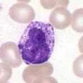
Wright's staining of multiple myeloma, plasmablastic type
-
Lymphocyte
-
 Basophil
 -
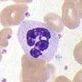 Neutrophil


Medical Disclaimer: WikiMD is for informational purposes only and is not a substitute for professional medical advice. Content may be inaccurate or outdated and should not be used for diagnosis or treatment. Always consult your healthcare provider for medical decisions. Verify information with trusted sources such as CDC.gov and NIH.gov. By using this site, you agree that WikiMD is not liable for any outcomes related to its content. See full disclaimer.
Credits:Most images are courtesy of Wikimedia commons, and templates, categories Wikipedia, licensed under CC BY SA or similar.
Translate page: - East Asian
中文,
日本,
한국어,
South Asian
हिन्दी,
தமிழ்,
తెలుగు,
Urdu,
ಕನ್ನಡ,
Southeast Asian
Indonesian,
Vietnamese,
Thai,
မြန်မာဘာသာ,
বাংলা
European
español,
Deutsch,
français,
Greek,
português do Brasil,
polski,
română,
русский,
Nederlands,
norsk,
svenska,
suomi,
Italian
Middle Eastern & African
عربى,
Turkish,
Persian,
Hebrew,
Afrikaans,
isiZulu,
Kiswahili,
Other
Bulgarian,
Hungarian,
Czech,
Swedish,
മലയാളം,
मराठी,
ਪੰਜਾਬੀ,
ગુજરાતી,
Portuguese,
Ukrainian

.jpg)