Abdominal ultrasonography
Abdominal ultrasonography is a medical imaging technique used to visualize the organs and structures in the abdomen. It employs ultrasound waves to produce images of the abdominal organs, including the liver, gallbladder, pancreas, spleen, kidneys, and bladder.
Technique[edit]
Abdominal ultrasonography is performed using a device called a transducer, which emits high-frequency sound waves. These sound waves penetrate the body and are reflected back to the transducer by the internal organs. The reflected waves are then converted into electrical signals, which are processed to form an image on a monitor.
The procedure is non-invasive and typically requires the patient to lie on their back. A water-based gel is applied to the skin to facilitate the transmission of sound waves. The transducer is moved over the abdomen to capture images from different angles.
Applications[edit]
Abdominal ultrasonography is used for a variety of diagnostic purposes, including:
- Evaluating abdominal pain
- Detecting gallstones
- Assessing liver disease
- Diagnosing kidney stones
- Monitoring the size and shape of abdominal organs
Advantages[edit]
Abdominal ultrasonography offers several advantages:
- It is a safe procedure with no exposure to ionizing radiation.
- It provides real-time imaging, allowing for dynamic assessment of organs.
- It is relatively inexpensive compared to other imaging modalities like CT scans or MRI.
Limitations[edit]
While abdominal ultrasonography is a valuable diagnostic tool, it has limitations:
- It may not provide clear images in patients with obesity or excessive intestinal gas.
- It is operator-dependent, requiring skill and experience to obtain accurate results.
Related Pages[edit]
Gallery[edit]
-
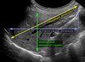
 Ultrasonographic measurement of aortic diameter
 -
 Liver measurements on ultrasonography
 -
 Morison's pouch on ultrasound
 -
Abdominal ultrasound full exam
-
Abdominal ultrasound full exam
-
Abdominal ultrasound full exam
-
Abdominal ultrasound full exam
-
Abdominal ultrasound full exam
-
Abdominal ultrasound full exam


Abdominal_ultrasonography[edit]

Medical Disclaimer: WikiMD is for informational purposes only and is not a substitute for professional medical advice. Content may be inaccurate or outdated and should not be used for diagnosis or treatment. Always consult your healthcare provider for medical decisions. Verify information with trusted sources such as CDC.gov and NIH.gov. By using this site, you agree that WikiMD is not liable for any outcomes related to its content. See full disclaimer.
Credits:Most images are courtesy of Wikimedia commons, and templates, categories Wikipedia, licensed under CC BY SA or similar.
Translate page: - East Asian
中文,
日本,
한국어,
South Asian
हिन्दी,
தமிழ்,
తెలుగు,
Urdu,
ಕನ್ನಡ,
Southeast Asian
Indonesian,
Vietnamese,
Thai,
မြန်မာဘာသာ,
বাংলা
European
español,
Deutsch,
français,
Greek,
português do Brasil,
polski,
română,
русский,
Nederlands,
norsk,
svenska,
suomi,
Italian
Middle Eastern & African
عربى,
Turkish,
Persian,
Hebrew,
Afrikaans,
isiZulu,
Kiswahili,
Other
Bulgarian,
Hungarian,
Czech,
Swedish,
മലയാളം,
मराठी,
ਪੰਜਾਬੀ,
ગુજરાતી,
Portuguese,
Ukrainian
