Electroretinography
Electroretinography (ERG) is a diagnostic test that measures the electrical activity generated by neural and non-neural cells in the retina in response to a visual stimulus. The retina is the light-sensitive tissue at the back of the eye, and its function is crucial for vision. ERG is an important tool in the diagnosis and management of various retinal diseases and conditions.
Overview[edit]
Electroretinography involves the use of electrodes placed on the surface of the eye or on the skin surrounding the eye to record electrical responses from the retina when it is stimulated by light. The test is non-invasive and can provide critical information about the function of the retina, which cannot be obtained through a standard eye examination.
Indications[edit]
ERG is indicated for the diagnosis and monitoring of a wide range of retinal conditions, including:
- Retinitis Pigmentosa and other forms of inherited retinal dystrophies
- Diabetic Retinopathy, to assess the extent of retinal damage
- Retinal Detachment, to evaluate retinal function
- Unexplained visual loss, when other diagnostic tests have failed to identify the cause
- Toxicity from medications known to affect retinal function, such as hydroxychloroquine
Procedure[edit]
The standard ERG procedure involves dilating the pupils with eye drops to allow for maximum light stimulation of the retina. The patient is then seated in a dark room to adapt their eyes to the darkness. A special contact lens electrode is placed on the cornea, and a ground electrode is attached to the skin. The patient is exposed to a series of light flashes, and the electrical responses of the retina are recorded. The procedure typically takes about an hour.
Types of ERG[edit]
There are several types of ERG, each designed to test different functions of the retina:
- Full-field ERG (ffERG): Records a global response from the entire retina.
- Multifocal ERG (mfERG): Provides detailed information about the function of the central retina.
- Pattern ERG (pERG): Measures the function of the macula, the central part of the retina responsible for detailed vision.
- Oscillatory Potentials (OPs): Assess the function of the inner retinal layers.
Interpretation[edit]
The results of an ERG are interpreted by comparing the amplitude (height) and timing (latency) of the recorded waveforms to normal values. Abnormal ERG results can indicate retinal dysfunction, which may be due to a variety of retinal diseases or conditions.
Risks and Complications[edit]
ERG is generally safe, with minimal risks. Patients may experience discomfort from the contact lens electrode, and the eye drops used to dilate the pupils can cause temporary blurred vision and sensitivity to light. There is a very low risk of infection from the contact lens.
Conclusion[edit]
Electroretinography is a valuable diagnostic tool in the field of ophthalmology, providing essential information about the functional status of the retina. It is used in the diagnosis and management of a wide range of retinal conditions, contributing to the preservation and improvement of vision in affected patients.
This medical article is a stub. You can help WikiMD by expanding the page. |
Electroretinography[edit]
-
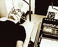
 Maximal Response ERG
 -
 Graph AB ERG Electroretinography
 -
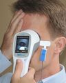 2014 ERG Test
 -
 Electroretinogram


Medical Disclaimer: WikiMD is for informational purposes only and is not a substitute for professional medical advice. Content may be inaccurate or outdated and should not be used for diagnosis or treatment. Always consult your healthcare provider for medical decisions. Verify information with trusted sources such as CDC.gov and NIH.gov. By using this site, you agree that WikiMD is not liable for any outcomes related to its content. See full disclaimer.
Credits:Most images are courtesy of Wikimedia commons, and templates, categories Wikipedia, licensed under CC BY SA or similar.
Translate page: - East Asian
中文,
日本,
한국어,
South Asian
हिन्दी,
தமிழ்,
తెలుగు,
Urdu,
ಕನ್ನಡ,
Southeast Asian
Indonesian,
Vietnamese,
Thai,
မြန်မာဘာသာ,
বাংলা
European
español,
Deutsch,
français,
Greek,
português do Brasil,
polski,
română,
русский,
Nederlands,
norsk,
svenska,
suomi,
Italian
Middle Eastern & African
عربى,
Turkish,
Persian,
Hebrew,
Afrikaans,
isiZulu,
Kiswahili,
Other
Bulgarian,
Hungarian,
Czech,
Swedish,
മലയാളം,
मराठी,
ਪੰਜਾਬੀ,
ગુજરાતી,
Portuguese,
Ukrainian
