Fluorescence imaging
Fluorescence Imaging is a technique used in biological research and medical diagnostics to visualize cellular and subcellular structures within living organisms. It uses the property of fluorescence, where certain compounds, known as fluorophores, emit light of a longer wavelength when they absorb light of a shorter wavelength.
History[edit]
The concept of fluorescence was first observed by Sir George Gabriel Stokes in 1852. However, the application of fluorescence in imaging was not realized until the 20th century, with the development of the fluorescence microscope.
Principles[edit]
Fluorescence imaging is based on the principle of fluorescence. When a fluorophore absorbs light of a specific wavelength, it gets excited to a higher energy state. After a short delay, it returns to its ground state by emitting light of a longer wavelength. This emitted light is then captured to form an image.
Techniques[edit]
There are several techniques in fluorescence imaging, including:
- Fluorescence microscopy: This is the most common technique, used to study the structure and function of cells.
- Confocal microscopy: This technique uses a pinhole to eliminate out-of-focus light, resulting in clearer images.
- Two-photon excitation microscopy: This technique uses two photons of lower energy to excite the fluorophore, allowing for deeper tissue imaging.
- Fluorescence resonance energy transfer (FRET): This technique is used to measure the distance between two fluorophores.
Applications[edit]
Fluorescence imaging has a wide range of applications in both research and clinical settings. It is used in cell biology to study the structure and function of cells, in molecular biology to study protein interactions, and in medical diagnostics to detect diseases such as cancer.
See also[edit]
- Fluorescence
- Fluorescence microscope
- Confocal microscopy
- Two-photon excitation microscopy
- Fluorescence resonance energy transfer (FRET)
References[edit]
This medical imaging related article is a stub. You can help WikiMD by expanding it.
-
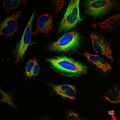 Multicolor fluorescence image of living HeLa cells
 -
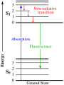 Jablonski Diagram of Fluorescence
 -
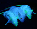 GFP Mice
 -
Agarose gel with UV illumination - Ethidium bromide stained DNA glows orange
-
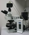 Olympus BX61 fluorescence microscope
 -
Fluorescence from Fluorescent Proteins


Medical Disclaimer: WikiMD is for informational purposes only and is not a substitute for professional medical advice. Content may be inaccurate or outdated and should not be used for diagnosis or treatment. Always consult your healthcare provider for medical decisions. Verify information with trusted sources such as CDC.gov and NIH.gov. By using this site, you agree that WikiMD is not liable for any outcomes related to its content. See full disclaimer.
Credits:Most images are courtesy of Wikimedia commons, and templates, categories Wikipedia, licensed under CC BY SA or similar.
Translate page: - East Asian
中文,
日本,
한국어,
South Asian
हिन्दी,
தமிழ்,
తెలుగు,
Urdu,
ಕನ್ನಡ,
Southeast Asian
Indonesian,
Vietnamese,
Thai,
မြန်မာဘာသာ,
বাংলা
European
español,
Deutsch,
français,
Greek,
português do Brasil,
polski,
română,
русский,
Nederlands,
norsk,
svenska,
suomi,
Italian
Middle Eastern & African
عربى,
Turkish,
Persian,
Hebrew,
Afrikaans,
isiZulu,
Kiswahili,
Other
Bulgarian,
Hungarian,
Czech,
Swedish,
മലയാളം,
मराठी,
ਪੰਜਾਬੀ,
ગુજરાતી,
Portuguese,
Ukrainian

.jpg)