Lateral ventricles
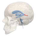
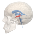
Lateral ventricles are structures within the brain that are part of the ventricular system. They are paired cavities, one in each cerebral hemisphere, and are the largest of the brain's ventricles. The lateral ventricles are filled with cerebrospinal fluid (CSF), which is produced by the choroid plexus located within these ventricles. CSF plays a crucial role in protecting the brain by providing a cushioning effect, removing waste products, and supplying nutrients to the central nervous system.
Structure[edit]
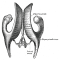
The lateral ventricles are divided into several parts: the frontal (anterior) horn, the body, the atrium (or trigone), the occipital (posterior) horn, and the temporal (inferior) horn. Each part is located within a specific region of the cerebral hemisphere. The frontal horn is situated in the frontal lobe, the body extends back into the parietal lobe, the atrium lies at the junction of the temporal, parietal, and occipital lobes, the occipital horn extends into the occipital lobe, and the temporal horn reaches into the temporal lobe.
Function[edit]
The primary function of the lateral ventricles is to produce and circulate cerebrospinal fluid. This fluid flows from the lateral ventricles through the interventricular foramina (or foramina of Monro) into the third ventricle, then into the fourth ventricle, and finally into the subarachnoid space surrounding the brain and spinal cord. CSF acts as a shock absorber, reducing the impact of trauma to the brain. It also helps in the regulation of intracranial pressure, provides a medium for the exchange of nutrients and waste products between the brain and blood, and creates a chemically stable environment for the brain.
Clinical Significance[edit]
Abnormalities in the size, shape, or function of the lateral ventricles can be indicative of various medical conditions. Hydrocephalus is a condition characterized by an excessive accumulation of CSF in the ventricles, leading to increased intracranial pressure and potentially causing damage to brain tissues. It can result from obstruction of CSF flow, overproduction of CSF, or impaired absorption of CSF. Other conditions related to the lateral ventricles include intraventricular hemorrhage, which is bleeding into the ventricles, and ventriculomegaly, which is an abnormal enlargement of the ventricles often associated with developmental anomalies or brain injuries.
Diagnosis and Treatment[edit]
Imaging techniques such as computed tomography (CT) scans and magnetic resonance imaging (MRI) are commonly used to assess the lateral ventricles for abnormalities. These imaging modalities can provide detailed information about the size, shape, and position of the ventricles, as well as any associated abnormalities of the brain.
Treatment of conditions affecting the lateral ventricles depends on the underlying cause. In cases of hydrocephalus, for example, treatment may involve the surgical insertion of a ventriculoperitoneal shunt to drain excess CSF from the brain to another part of the body where it can be absorbed. Alternatively, a procedure known as endoscopic third ventriculostomy (ETV) may be performed to create a new pathway for CSF flow, bypassing the obstruction.
Conclusion[edit]
The lateral ventricles play a vital role in the production and circulation of cerebrospinal fluid, essential for the protection and functioning of the brain. Understanding the structure, function, and clinical significance of these ventricles is crucial for diagnosing and treating conditions that affect the ventricular system and the brain as a whole.
This medical article is a stub. You can help WikiMD by expanding the page. |
-
 Lateral ventricles
 -
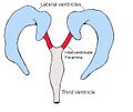 Interventricular foramina
 -
 Anterior horn of lateral ventricle
 -
 Body of lateral ventricle
 -
 Trigone of lateral ventricle
 -
 Posterior horn of lateral ventricle
 -
 Inferior horn of lateral ventricle
 -
 Lateral ventricles
 -
 Lateral ventricles


Medical Disclaimer: WikiMD is for informational purposes only and is not a substitute for professional medical advice. Content may be inaccurate or outdated and should not be used for diagnosis or treatment. Always consult your healthcare provider for medical decisions. Verify information with trusted sources such as CDC.gov and NIH.gov. By using this site, you agree that WikiMD is not liable for any outcomes related to its content. See full disclaimer.
Credits:Most images are courtesy of Wikimedia commons, and templates, categories Wikipedia, licensed under CC BY SA or similar.
Translate page: - East Asian
中文,
日本,
한국어,
South Asian
हिन्दी,
தமிழ்,
తెలుగు,
Urdu,
ಕನ್ನಡ,
Southeast Asian
Indonesian,
Vietnamese,
Thai,
မြန်မာဘာသာ,
বাংলা
European
español,
Deutsch,
français,
Greek,
português do Brasil,
polski,
română,
русский,
Nederlands,
norsk,
svenska,
suomi,
Italian
Middle Eastern & African
عربى,
Turkish,
Persian,
Hebrew,
Afrikaans,
isiZulu,
Kiswahili,
Other
Bulgarian,
Hungarian,
Czech,
Swedish,
മലയാളം,
मराठी,
ਪੰਜਾਬੀ,
ગુજરાતી,
Portuguese,
Ukrainian
