Mammary gland
(MA-muh-ree ...) Glandular organ located on the chest. The mammary gland is made up of connective tissue, fat, and tissue that contains the glands that can make milk. Also called breast.
Anatomy > Gray's Anatomy of the Human Body > XI. Splanchnology > 3d. 6. The Mammæ
Henry Gray (1821–1865). Anatomy of the Human Body. 1918. 3d. 6. The Mammæ Breasts
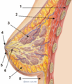
The mammæ secrete the milk, and are accessory glands of the generative system. They exist in the male as well as in the female; but in the former only in the rudimentary state, unless their growth is excited by peculiar circumstances. In the female they are two large hemispherical eminences lying within the superficial fascia and situated on the front and sides of the chest; each extends from the second rib above to the sixth rib below, and from the side of the sternum to near the midaxillary line. Their weight and dimensions differ at different periods of life, and in different individuals. Before puberty they are of small size, but enlarge as the generative organs become more completely developed. They increase during pregnancy and especially after delivery, and become atrophied in old age. The left mamma is generally a little larger than the right. The deep surface of each is nearly circular, flattened, or slightly concave, and has its long diameter directed upward and lateralward toward the axilla; it is separated from the fascia covering the Pectoralis major, Serratus anterior, and Obliquus externus abdominis by loose connective tissue. The subcutaneous surface of the mamma is convex, and presents, just below the center, a small conical prominence, the papilla
The Mammary Papilla or Nipple (papilla mammæ) is a cylindrical or conical eminence situated about the level of the fourth intercostal space. It is capable of undergoing a sort of erection from mechanical excitement, a change mainly due to the contraction of its muscular fibers. It is of a pink or brownish hue, its surface wrinkled and provided with secondary papillæ; and it is perforated by from fifteen to twenty orifices, the apertures of the lactiferous ducts.
The base of the mammary papilla is surrounded by an areola In the virgin the areola is of a delicate rosy hue; about the second month after impregnation it enlarges and acquires a darker tinge, and as pregnancy advances it may assume a dark brown or even black color. This color diminishes as soon as lactation is over, but is never entirely lost throughout life. These changes in the color of the areola are of importance in forming a conclusion in a case of suspected first pregnancy. Near the base of the papilla, and upon the surface of the areola, are numerous large sebaceous glands, the areolar glands which become much enlarged during lactation, and present the appearance of small tubercles beneath the skin. These glands secrete a peculiar fatty substance, which serves as a protection to the integument of the papilla during the act of sucking.
The mammary papilla consists of numerous vessels, intermixed with plain muscular fibers, which are principally arranged in a circular manner around the base: some few fibers radiating from base to apex.
Development—The mamma is developed partly from mesoderm and partly from ectoderm—its bloodvessels and connective tissue being derived from the former, its cellular elements from the latter. Its first rudiment is seen about the third month, in the form of a number of small inward projections of the ectoderm, which invade the mesoderm; from these, secondary tracts of cellular elements radiate and subsequently give rise to the epithelium of the glandular follicles and ducts. The development of the follicles, however, remains imperfect, except in the parous female. Structure (Figs. 1172, 1173)—The mamma consists of gland tissue; of fibrous tissue, connecting its lobes; and of fatty tissue in the intervals between the lobes. The gland tissue, when freed from fibrous tissue and fat, is of a pale reddish color, firm in texture, flattened from before backward and thicker in the center than at the circumference.
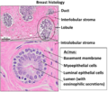
The subcutaneous surface of the mamma presents numerous irregular processes which project toward the skin and are joined to it by bands of connective tissue. It consists of numerous lobes, and these are composed of lobules, connected together by areolar tissue, bloodvessels, and ducts. The smallest lobules consist of a cluster of rounded alveoli, which open into the smallest branches of the lactiferous ducts; these ducts unite to form larger ducts, and these end in a single canal, corresponding with one of the chief subdivisions of the gland. The number of excretory ducts varies from fifteen to twenty; they are termed the tubuli lactiferi They converge toward the areola, beneath which they form dilatations or ampullæ which serve as reservoirs for the milk, and, at the base of the papillæ, become contracted, and pursue a straight course to its summit, perforating it by separate orifices considerably narrower than the ducts themselves.
The ducts are composed of areolar tissue containing longitudinal and transverse elastic fibers; muscular fibers are entirely absent; they are lined by columnar epithelium resting on a basement membrane. The epithelium of the mamma differs according to the state of activity of the organ. In the gland of a woman who is not pregnant or suckling, the alveoli are very small and solid, being filled with a mass of granular polyhedral cells. During pregnancy the alveoli enlarge, and the cells undergo rapid multiplication. At the commencement of lactation, the cells in the center of the alveolus undergo fatty degeneration, and are eliminated in the first milk, as colostrum corpuscles The peripheral cells of the alveolus remain, and form a single layer of granular, short columnar cells, with spherical nuclei, lining the basement membrane. The cells, during the state of activity of the gland, are capable of forming, in their interior, oil globules, which are then ejected into the lumen of the alveolus, and constitute the milk globules. When the acini are distended by the accumulation of the secretion the lining epithelium becomes flattened.
FIG. 1172– Dissection of the lower half of the mamma during the period of lactation. (Luschka.) (Picture From the Classic Gray's Anatomy)
FIG. 1173– Section of portion of mamma. (Picture From the Classic Gray's Anatomy) The fibrous tissue invests the entire surface of the mamma, and sends down septa between its lobes, connecting them together. The fatty tissue covers the surface of the gland, and occupies the interval between its lobes. It usually exists in considerable abundance, and determines the form and size of the gland. There is no fat immediately beneath the areola and papilla. Vessels and Nerves—The arteries supplying the mammæ are derived from the thoracic branches of the axillary, the intercostals, and the internal mammary. The veins describe an anastomotic circle around the base of the papilla, called by Haller the circulus venosus From this, large branches transmit the blood to the circumference of the gland, and end in the axillary and internal mammary veins. The lymphatics are described on page 715. The nerves are derived from the anterior and lateral cutaneous branches of the fourth, fifth, and sixth thoracic nerves.
Gray's Anatomy Contents | Gray's Anatomy Subject Index[edit]
| Anatomy of the breast | ||||
|---|---|---|---|---|
|
-
 Mammary gland
 -
 Mammary gland
 -
Mammary gland
-
Mammary gland
-
Mammary gland
-
Mammary gland
-
 Mammary gland
 -
Mammary gland
-
Mammary gland
-
Mammary gland
-
Mammary gland


Medical Disclaimer: WikiMD is for informational purposes only and is not a substitute for professional medical advice. Content may be inaccurate or outdated and should not be used for diagnosis or treatment. Always consult your healthcare provider for medical decisions. Verify information with trusted sources such as CDC.gov and NIH.gov. By using this site, you agree that WikiMD is not liable for any outcomes related to its content. See full disclaimer.
Credits:Most images are courtesy of Wikimedia commons, and templates, categories Wikipedia, licensed under CC BY SA or similar.
Translate page: - East Asian
中文,
日本,
한국어,
South Asian
हिन्दी,
தமிழ்,
తెలుగు,
Urdu,
ಕನ್ನಡ,
Southeast Asian
Indonesian,
Vietnamese,
Thai,
မြန်မာဘာသာ,
বাংলা
European
español,
Deutsch,
français,
Greek,
português do Brasil,
polski,
română,
русский,
Nederlands,
norsk,
svenska,
suomi,
Italian
Middle Eastern & African
عربى,
Turkish,
Persian,
Hebrew,
Afrikaans,
isiZulu,
Kiswahili,
Other
Bulgarian,
Hungarian,
Czech,
Swedish,
മലയാളം,
मराठी,
ਪੰਜਾਬੀ,
ગુજરાતી,
Portuguese,
Ukrainian
